.png)


Why Imaging Matters in Modern GI Oncology
Advanced imaging underpins every step of gastrointestinal cancer care. High‑resolution CT, MRI, and PET/CT define tumor size, nodal involvement, and distant metastases, enabling accurate staging and prognostication. These datasets feed into image‑guided radiation planning, where soft‑tissue contrast and functional maps guide target delineation, organ‑at‑risk sparing, and dose‑painting strategies. Seamless integration of imaging findings into multidisciplinary tumor boards ensures surgeons, medical oncologists, and radiologists coordinate personalized treatment plans, optimizing curative intent while minimizing toxicity.
Imaging Foundations for GI Radiotherapy

Accurate imaging underpins every step of gastrointestinal (GI) radiotherapy, from initial staging to daily‑guided delivery. Computed tomography (CT) and four‑dimensional CT (4D‑CT) supply rapid, three‑dimensional anatomy and capture respiratory motion, enabling creation of internal target volumes that protect moving organs such as the pancreas and liver. Magnetic resonance imaging (MRI), especially multiparametric MRI, delivers superior soft‑tissue contrast and functional data (diffusion‑weighted, dynamic contrast‑enhanced) that delineate tumor borders and identify high‑risk sub‑volumes for dose painting. Positron emission tomography (PET) and PET/CT reveal metabolic activity, highlighting aggressive disease and occult metastases, which guides biologically‑adaptive planning. Ultrasound, particularly endoscopic ultrasound (EUS) provides high‑resolution, real‑time images for tumor assessment and biopsy, and can be fused with CT or MRI for precise target definition. Cone‑beam CT (CBCT) integrated into linear accelerators enables image‑guided radiotherapy (IGRT), verifying patient positioning and anatomy before each fraction and supporting online adaptive radiotherapy.
Diagnostic imaging techniques such as multiphasic contrast‑enhanced CT, MRI, MRICP, PET/CT, and EUS together allow multidisciplinary teams to tailor pancreatic cancer treatment with confidence.
The four primary medical‑imaging modalities are X‑ray, CT, MRI, and ultrasound each offering unique strengths for anatomic and functional assessment.
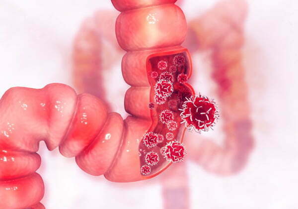
Radiology employs X‑ray, CT, MRI, ultrasound, and nuclear scans like PET to visualize pathology, guide biopsies, and monitor treatment response across the GI tract.
Adaptive Radiotherapy: Merging Imaging with Real‑Time Planning

Radiology procedures – Radiology procedures encompass a broad range of imaging‑guided diagnostic and therapeutic techniques that allow physicians to visualize internal structures and, when needed, intervene without open surgery. Common diagnostic studies include X‑ray radiography, CT scans, MRI, mammography, bone‑density (DEXA) scans, and nuclear‑medicine PET scans, each providing detailed pictures of organs, vessels, and tissues. Interventional radiology adds minimally invasive treatments such as CT‑ or ultrasound‑guided biopsies, tumor ablations, embolizations, drainage placements, and catheter‑based therapies, all performed under real‑time imaging guidance. These procedures are essential in oncology for accurately staging cancer, planning surgery, delivering targeted therapies, and monitoring treatment response while minimizing patient discomfort and recovery time. At Hirschfeld Oncology, our radiology team works closely with oncologists to ensure that every imaging and image‑guided intervention is tailored to each patient’s cancer care plan.
Interventional Radiology – Interventional radiology (IR) is a minimally invasive, imaging‑guided specialty that diagnoses and treats disease by inserting catheters, needles, or other devices through the skin. Using X‑ray, CT, ultrasound, or MRI for guidance, IR can perform procedures such as tumor biopsies, embolization, radiofrequency or microwave ablation, and placement of vascular stents or chemotherapy ports—often avoiding the need for open surgery. In pancreatic cancer, IR is frequently used to relieve biliary obstruction with stents, deliver localized ablative therapy, and obtain tissue samples for precise diagnosis. These image‑guided interventions provide faster recovery, less pain, and reduced hospital stays while complementing systemic chemotherapy and radiation. At Hirschfeld Oncology, interventional radiologists collaborate with surgeons, medical oncologists, and supportive‑care teams to integrate these targeted treatments into a comprehensive, patient‑centered cancer‑care plan.
Clinical Impact: Outcomes, Toxicity, and Survival

Recent adaptive radiotherapy (ART) data underscore its value in gastrointestinal (GI) malignancies. The multicenter SMART phase‑II trial of MRgRT‑SBRT for borderline‑resectable or locally advanced pancreatic cancer (136 patients) reported 0 % acute GI toxicity, a 78.2 % two‑year local‑control rate, and a median overall survival of 22.9 months. Ongoing ARTIA‑pancreas ([CTgART](./adaptive-radiotherapy-real-time-treatment-adjustments-for-pancreatic-cancer#the-patient-experience-what-to-expect-from-an-adaptive-treatment)) and NRG GI‑011 (CTgART trials are evaluating whether similar dose‑escalation can be achieved with comparable toxicity profiles. Dose escalation to ablative regimens—e.g., 50 Gy in five fractions (BED ≈ 100 Gy)—has improved local control for pancreatic and primary liver cancers, while online adaptation spares adjacent OARs such as duodenum, small bowel, and stomach. In liver SBRT, 53 % of 99 patients required plan adaptation, yielding 91 % one‑year local control and low high‑grade GI toxicity. Survival outcomes vary by disease site: pancreatic cancer median OS with ART‑guided SBRT is ≈23 months, whereas 5‑year survival for localized stomach cancer approaches 75 % and drops to <8 % with distant metastasis.
Early symptoms of gastrointestinal cancer
Persistent abdominal pain, new‑onset heartburn, early satiety, unexplained weight loss, anemia, and occult GI bleeding are common early signs that merit prompt evaluation.
Gastrointestinal cancer survival rate
5‑year relative survival is ≈36‑38 % overall; localized gastric disease reaches ≈75‑77 %, regional disease ≈35‑37 %, and metastatic disease ≈7‑8 %.
Radiation therapy side effects
Acute effects include fatigue, skin erythema, nausea, vomiting, and organ‑specific irritation; late effects depend on dose, field, and patient factors.
Personalizing Care: Genomics, Radiomics, and AI

Radiomics and machine‑learning models extract quantitative texture features from CT, MRI, and PET scans, enabling prediction of radiosensitivity and toxicity in GI malignancies. These data feed into genomic‑adjusted radiation dose (GARD), which multiplies prescribed dose by a tumor‑specific radiation‑sensitivity index derived from a 10‑gene expression panel, allowing dose escalation for resistant tumors while sparing normal tissue. AI‑driven auto‑segmentation dramatically speeds up contouring of gross tumor volumes and organs‑at‑risk; synthetic CT generation from MRI or CBCT further streamlines MR‑guided adaptive workflows. MR elastography adds a stiffness map that highlights desmoplastic, potentially radio‑resistant sub‑volumes, guiding focal dose boosts.
Gastrointestinal cancer causes: Combination of modifiable lifestyle factors (obesity, sedentary behavior, high‑fat processed foods, alcohol, tobacco) and non‑modifiable biological influences (chronic diseases, inherited mutations, microbiota alterations) drives GI tumor development.
Most common GI cancer: Colorectal cancer is the leading GI malignancy in the United States, arising from adenomatous polyps and affecting primarily adults over 50, with rising incidence in younger cohorts.
Medical imaging techniques PDF: Such a PDF outlines physical principles, clinical applications, and comparative strengths of X‑ray, CT, MRI, ultrasound, and nuclear medicine, includes historical milestones, sample images, and discusses emerging AI‑enhanced analysis for cancer diagnosis and treatment planning.
Future Directions: Emerging Modalities and Clinical Trials

Advanced imaging is reshaping gastrointestinal (GI) cancer therapy. MR‑Linac platforms now enable real‑time MR‑guided SBRT, allowing daily re‑contouring and dose escalation while sparing duodenum, stomach and bowel—crucial for pancreatic and liver lesions. Hybrid PET/MRI and functional imaging (DW‑MRI, DCE‑MRI) support dose‑painting by identifying high‑metabolic or hypoxic sub‑volumes for selective boost. Magnetic resonance elastography (MRE) adds a stiffness map; stiff, potentially radio‑resistant regions can receive intensified dose without exceeding OAR constraints. Direct‑to‑unit (DTU) workflows combine diagnostic imaging with online ART, eliminating a separate CT simulation and, coupled with AI‑driven auto‑contouring and auto‑planning, compress the 40‑90 minute per‑fraction burden to minutes. Ongoing trials are testing these concepts: ARTIA‑pancreas (CTgART), NRG GI‑011 LAP‑100 (CTgART/MRgART), and SMART ONE (single‑fraction SBRT for abdominal oligometastases).
Gastrointestinal cancer treatment – At Hirschfeld Oncology, care is multidisciplinary and personalized, integrating surgery, systemic therapy, modern IMRT/SBRT, and adaptive plans based on daily imaging and biomarker data.
Gastrointestinal cancer how to detect – Detection begins with history, physical exam, and risk‑factor assessment, followed by contrast‑enhanced CT, MRI or ultrasound for anatomic staging, endoscopic visualization with biopsy for histology, and adjunctive PET/CT for metabolic mapping.
Brachytherapy Radiology – Brachytherapy uses imaging‑guided placement of radioactive seeds or catheters (CT, MRI, fluoroscopy, ultrasound) to deliver high‑dose radiation directly to the tumor while sparing surrounding tissue, offering a precise, minimally invasive option for select GI lesions.
A New Era of Imaging‑Driven Radiation Oncology
The integration of multiparametric MRI, PET/CT, and AI‑driven auto‑segmentation with daily cone‑beam or MR‑Linac imaging enables true adaptive radiotherapy, tailoring dose to each patient’s anatomy and biology. At Hirschfeld Oncology this imaging‑driven approach translates into higher local control, reduced gastrointestinal toxicity, and personalized treatment pathways. Ongoing participation in trials such as SMART, ARTIA‑pancreas, and NRG‑GI‑011 ensures rapid adoption of novel dose‑painting, proton‑based, and biologically‑guided strategies, continually advancing care for gastrointestinal malignancies for the future of GI cancer care and research.
.png)
.png)