.png)


Receiving a notice for a callback mammogram can be unsettling, but it’s important to know what it really means. Simply put, the radiologist who read your images saw something that needs a closer look.
It’s not a diagnosis. It's just a request for more information. I often tell my patients to think of it like a photographer taking a group photo. Sometimes, one person is a little blurry, so the photographer takes a few extra, more focused shots of that person to make sure the final picture is perfect. That's exactly what's happening here.
Demystifying the Mammogram Callback Notice
The moment that phone rings or you open that letter, it's completely natural for your mind to jump to the worst-case scenario. Take a deep breath. A callback is a very routine part of the screening process, designed to make sure your results are as accurate as possible. The main goal is simply to clear up any uncertainty from the first set of images.
Callbacks are surprisingly common, happening for about 10% of women who get a screening mammogram. That number is often a bit higher if it's your very first one. Without any previous mammograms to compare to, radiologists are extra careful to investigate anything that looks even slightly unusual.
Think of your first mammogram as the first "fingerprint" of your breast tissue. Radiologists don't yet know what's normal for you, so they may call you back to confirm that a specific finding is just part of your unique anatomy.
This cautious approach is actually a sign of a high-quality screening program. The vast majority of the time, this extra look confirms that everything is perfectly fine and the area in question is benign (not cancerous).
Putting the Statistics into Perspective
Let’s look at the numbers, because they can be incredibly reassuring. While it’s true that about 1 in 10 women will be called back after a screening mammogram, the story doesn't end there.
- Of all the women called back for more imaging, only about 1 in 5 will actually need a biopsy.
- And for those who do have a biopsy, 80% of the results come back as benign.
When you do the math, it means that only a very small percentage of women who get that callback notice—around 5%—are ultimately diagnosed with breast cancer. To put it another way, for every 100 women called back for a closer look, 95 of them will get the all-clear.
The Role of the Radiologist
A radiologist's job is to be a meticulous detective. They are trained to scrutinize every detail and spot the most subtle changes in breast tissue. If an area is hard to see clearly because of overlapping tissue, dense breasts, or even a technical glitch with the first image, they will always err on the side of caution.
Requesting more views is simply the next step in their diligent process. It's how they ensure nothing is missed, giving you the most accurate results and, ultimately, peace of mind.
Receiving a notice for a callback after your mammogram can be unsettling, but it helps to know what’s usually behind it. Most of the time, it’s a simple case of needing a clearer picture. Radiologists are trained to be meticulous, and if anything on your scan looks even slightly unclear, they’ll ask for more imaging to be absolutely certain.
This is a very common part of the breast screening process. It doesn't automatically mean something is wrong; it just means your care team is being extra careful. Let's walk through some of the most common, and often harmless, reasons you might get that call.
Common Reasons for a Mammogram Callback
To give you a quick overview, here are the most frequent reasons a radiologist might want to take a second look.
As you can see, the vast majority of callbacks are for reasons that have nothing to do with cancer. They are simply a way to ensure every screening is as accurate as possible.
Your First Mammogram Sets the Baseline
If this was your first time getting a mammogram, your chances of getting a callback are a little higher. Think of it this way: your breast tissue is like a unique fingerprint. Without any past images for comparison, the radiologist has no way of knowing what’s normal for you.
An area that might look suspicious could just be your unique anatomy. The follow-up images help them create a clear baseline, a detailed map of your breast tissue that will serve as the reference point for all your future screenings. This actually makes it much easier to spot real changes down the line.
Dense Breast Tissue Can Obscure the View
One of the most frequent reasons for a callback is dense breast tissue. It’s incredibly common—about 50% of women have it, and it’s perfectly normal. The issue is that on a mammogram, dense tissue shows up as white, and so do tumors or other areas of concern.
It's a bit like trying to spot a white cat in a snowstorm. The dense tissue can sometimes mask what lies beneath, making it tough for the radiologist to get a clear, confident read.
Follow-up imaging, like a breast ultrasound, is fantastic for this. It allows the radiologist to easily distinguish between normal dense tissue and something that needs more attention. Understanding how your family history of breast cancer might interact with factors like breast density can also give you a more complete view of your overall health profile.
Benign Findings That Need a Closer Look
Sometimes, a mammogram picks up something that is almost always benign (non-cancerous) but still needs to be confirmed. These common findings are a big reason for callbacks.
- Cysts: These are just simple, fluid-filled sacs. They are extremely common and are never cancerous. An ultrasound can confirm a finding is a cyst in a matter of minutes.
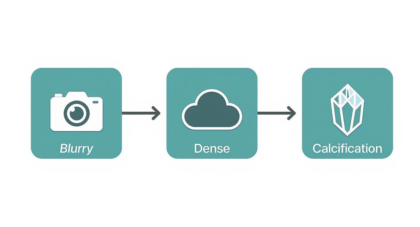
- Calcifications: These are tiny specks of calcium that can form in breast tissue. Most are harmless, but certain patterns—like a tight cluster—can sometimes be linked to very early cell changes. A magnified view helps the radiologist be sure.
- Asymmetry: It’s very normal for one breast to be shaped or structured a little differently than the other. A callback simply ensures this difference is part of your normal anatomy and not a new development.
Technical Issues and Image Quality
Finally, the reason for a callback might have nothing to do with your breasts at all. Just like any photograph, a mammogram needs to be perfectly clear to be useful.
A few technical hiccups can require a retake:
- Motion Blur: If you moved even a little during the scan, it could make the image blurry.
- Positioning: Sometimes, a small part of the breast tissue wasn't fully included in the first set of images.
- Artifacts: Believe it or not, deodorant, lotion, or body powder can show up on a mammogram as tiny white specks that look just like calcifications.
It’s also helpful to know that callback rates are closely tracked. In the U.S., the average recall rate is about 10.6%. Interestingly, technology plays a role. 3D mammography (tomosynthesis) has a lower callback rate of 9.4% compared to 10.8% for standard 2D exams, because it gives radiologists a much clearer view from the start. You can explore the full research on these callback rates to get a deeper understanding of the data.
Navigating Your Follow-Up Appointment
Learning you need extra imaging after a call back mammogram can feel unsettling. Having a clear roadmap for the day helps turn that uncertainty into something manageable.
Instead of retaking the entire breast image, this visit zeroes in on the exact spot that raised questions. Think of your screening mammogram as a bird’s-eye view of a forest; now you’re sending in a scout with binoculars and a high-resolution camera to inspect that one suspicious tree.
What To Expect On The Day Of Your Appointment
The check-in process will look familiar—you’ll change into a gown and settle in on the imaging table—but you’ll notice a few key differences once the focused work begins.
- Appointment Length: Plan for about 1–2 Hours, since your team will perform extra views and often review images with you before you leave.
- Wear A Two-Piece Outfit: A top and bottom makes it easy to undress from the waist up without hassle.
- Skip Deodorant & Lotions: Powders, creams or sprays on your underarms or chest can leave artifacts on your images.
The Tools Used For A Closer Look
Your technologist and radiologist may use one or both of these techniques to get the clearest possible view:
- Diagnostic Mammogram: Rather than a broad survey, this captures targeted images of the area in question. Special paddles provide magnified views or spot compression, gently flattening a small section to spread out tissue and clarify shapes.
- Breast Ultrasound: Using sound waves instead of radiation, ultrasound excels at distinguishing solid masses from fluid-filled cysts. A warm gel and handheld transducer glide over your skin—painless, quick, and especially useful for dense breast tissue.
The Role Of The Radiologist During Your Visit
A diagnostic workup brings your radiologist into the room from start to finish. They review each new image in real time, so any decision—like adding an ultrasound—happens immediately.
Often, the radiologist will step in front of the monitor to walk you through what they see. That on-the-spot dialogue turns a day of waiting into a day of understanding.
Having the radiologist present to provide preliminary feedback can transform a day of anxiety into a day of clear answers.
By the end of your appointment, you’ll know whether the finding is benign, if a short-term follow-up (in 6 months) makes sense for extra caution, or if a biopsy is the next step for a definitive diagnosis.
Understanding Your Diagnostic Results
When your follow-up images are reviewed, the radiologist will place the finding into one of three categories. You might receive a fully benign result, which means you simply return to your normal screening schedule. Or, you could see a “likely benign” note, prompting a short-term check in six months. Finally, if something looks suspicious, a biopsy will be recommended to get a definitive answer.
In a benign outcome, nothing concerning appears. You’ll breathe easier and plan for your next annual exam.
With a likely benign finding, your care team opts for caution. They’ll ask you to come back in six months to make sure nothing changes—catching any subtle shifts early, without rushing into invasive procedures.
If images still raise questions, a biopsy is the next step. A small needle collects a tissue sample, and lab analysis provides the conclusive diagnosis.
- Benign Outcome: No extra tests until your routine screening.
- Likely Benign Follow-Up: A six-month check confirms stability.
- Biopsy Recommendation: Needle sampling delivers a clear result.
Key Insight Only about 5–10% of callbacks lead to a cancer diagnosis, which means more than 90% turn out to be false alarms. Keeping this in mind can help you stay calm and focused on next steps.
Three Possible Outcomes
A benign result is by far the most common pathway. Nearly 80% of biopsies after callbacks end up showing no cancer, so returning to annual screening is the outcome for most patients.
Likely benign findings add a safety layer. Radiologists revisit your images six months later and only move forward with a biopsy if growth or new suspicious features appear. This approach minimizes unnecessary procedures.
About 1.6% of patients receive a biopsy recommendation when recall rates stay within the 7%–9% sweet spot. That balance maximizes cancer detection while keeping extra biopsies low. Learn more on PMC (https://pmc.ncbi.nlm.nih.gov/articles/PMC8087168/).
What To Expect After Results
If a biopsy is recommended, you’ll receive clear scheduling instructions. The procedure usually takes 15–30 minutes under local anesthesia, and you’ll head home the same day. Expect pathology results within 3–7 business days, so you can discuss next steps promptly.
While waiting, consider these questions for your provider:
- What did the imaging show?
- How long until I get the biopsy results?
- Who will guide me through the next steps?
For more on treatment options and expert support, explore our guide on breast cancer care at Hirschfeld Oncology.
Remember, it’s normal to feel anxious. Lean on friends, join a support group, or speak with a counselor. Your care team is thorough, attentive, and here to help every step of the way.
Expert Support And Consultation
If you’d like personalized guidance, schedule a consultation at Hirschfeld Oncology in Brooklyn. Our board-certified oncologists and patient navigators are ready to support you from diagnosis through treatment.
Local resources include:
- Brooklyn Breast Cancer Network
- NYC American Cancer Society Helpline
- Bushwick Patient Navigator Program
Most callbacks end with benign results, reflecting a screening process designed with your well-being in mind. Trust your team, ask questions, and know you’re never alone on this journey.
What To Expect If A Biopsy Is Recommended

Getting a biopsy recommendation can feel unnerving. Yet, a core needle biopsy is a quick outpatient procedure that usually causes less discomfort than most people imagine.
In minutes, a local anesthetic numbs the spot. Then, using ultrasound or mammogram guidance, your radiologist pinpoints the exact area—think of it like GPS for your breast tissue.
Core Needle Biopsy Procedure
Check-In and Prep
You’ll change into a gown while the technologist marks the precise location.Numbing Injection
A quick pinch delivers local anesthesia, and the area goes numb in seconds.Tissue Sampling
Guided by live imaging, the radiologist gently collects several small cores of tissue.Bandaging and Recovery
A small bandage covers the site. After a brief rest, you can head home.
Expert Insight
“A core needle biopsy is like pulling a small soil sample,” explains Dr. Azriel Hirschfeld. “It gives a clear snapshot of what’s happening below the surface.”
Understanding Imaging Guidance
Real-time imaging tracks every move of the needle, transforming a blind poke into a precise procedure.
- Ultrasound Guidance: Uses sound waves for live visualization.
- Stereotactic Guidance: Combines two X-ray angles for exact targeting.
Technologists fine-tune settings to keep radiation low and images crisp, always prioritizing your comfort.
What You Might Feel
- Mild Pressure: You’ll notice gentle pushing as the needle enters.
- Brief Discomfort: Most people compare it to a routine blood draw.
- Aching Afterward: Some tenderness or soreness for 24–48 hours is normal.
Aftercare Tips
- Ice Packs: Apply for five minutes on, five minutes off to reduce swelling.
- Rest: Avoid heavy lifting or strenuous activity for a day or two.
- Monitor for Signs: Watch for redness, fever, or increased pain.
Timeline For Results
Your clinic will give you a clear schedule. Always ask: “When can I expect the results?”
Emotional Support And Questions To Ask
Waiting for pathology can stir up a lot of anxiety. Reach out to a friend, family member, or a support group—you don’t have to go through this alone.
- How many cores were taken?
- Will I receive a phone update?
- What happens if the result is benign?
Key Takeaway
Less than 15% of core needle biopsies confirm malignancy, meaning most findings are benign.
You’ll leave with written aftercare instructions and after-hours contact information. If possible, bring someone with you for support.
Request a consultation at Hirschfeld Oncology on their website for personalized guidance.
Your care team will walk you through every detail. With clear information, you can face this next phase with confidence.
If malignancy is confirmed, you’ll discuss options from surgery to targeted therapies. Hirschfeld Oncology integrates supportive care to focus on your comfort and well-being at every step.
Coping With The Stress Of A Callback
Waiting for a callback after a mammogram can feel like watching an oven timer slowly tick down on a cake you’ve never baked before. Your mind races through every “what if” scenario. Acknowledging that swirl of emotions is the first step in finding calm.
Sometimes, simple actions can cut through the anxiety:
- Focus on your breath for two minutes of mindfulness.
- Call a trusted friend or family member who will listen without judgment.
- Resist the urge to dive into endless web searches—they often fuel worst-case thoughts.
“A callback is a normal checkpoint in breast screening.”
Strategies To Manage Anxiety
Even a few moments of guided meditation can lower stress hormones and settle a racing mind. Stepping outside for a short walk or trying gentle yoga stretches offers both mental and physical relief.
Keeping a small journal helps you track patterns in your thoughts. Write one feeling per entry—this creates structure and makes emotions feel less overwhelming. You might also make simple coping cards with prompts like “I am strong” or “This will pass.”
If you find self-care isn’t enough, consider talking with a counselor or joining a stress-management workshop. And remember to lean on your healthcare team—radiologists and nurse navigators at Hirschfeld Oncology are there to answer questions and guide you through each step.
One large study of 3.5 million screening mammograms found that 76% of women returned for routine exams within 9–30 months. Those who experienced a false-positive result, however, were less likely to come back—only 61% did after diagnostic follow-up and 67% after biopsies. Learn more about these findings.
Building Confidence For Future Screenings
You don’t have to face this alone. Hirschfeld Oncology offers patient navigation and virtual support for Brooklyn residents. Inviting a friend can turn an anxious appointment into a shared experience.
Try these ideas to reclaim the narrative:
- Treat your mammogram as a self-care appointment.
- Pack noise-canceling headphones to block out distractions.
- Give yourself a small reward afterward—whether it’s tea or a walk in the park.
- Reach out to our patient navigators for tailored guidance.
Our team at Hirschfeld Oncology is here at every twist and turn. Remember, every callback is a step toward clarity.
Frequently Asked Questions About Mammogram Callbacks
It’s normal to have a few questions after that callback notice hits your mailbox. This quick guide breaks down timing, risks, and practical steps so you can feel prepared.
Key Takeaways
- Appointment Window: Follow-up imaging usually occurs within two weeks of your callback letter.
- Diagnosis Odds: Only about 5–10% of callbacks lead to a cancer diagnosis.
- Preparation Tips: Skip deodorant, wear a comfortable top, and try to stay still during photos for the clearest results.
How Long Will I Wait for Follow-Up and Results
Most imaging centers will slot you in within days, but it can stretch to two weeks. You’ll often get preliminary feedback during your diagnostic visit itself. If they recommend a biopsy, you can expect pathology results in about 3–7 business days, though you should always double-check the timeline with your clinic.
“Scheduling follow-up appointments promptly can reduce anxiety and prevent delays,” explains Dr. Hirschfeld.
Does a Callback Increase My Cancer Risk
Getting asked back doesn’t change your long-term risk. In the vast majority of cases, callbacks happen because of benign quirks on your image or simply image-quality checks. Sticking to your yearly mammogram schedule remains your best strategy for spotting any changes early.
For more in-depth answers, check out our detailed FAQs and additional resources at Hirschfeld Oncology FAQs and Resources.
Can I Prevent a Future Callback
Some variables—like dense breast tissue—are out of our hands. Still, there are simple habits that help:
- Wear no lotions, powders, or deodorants under your arms
- Dress in a two-piece outfit to make the process smoother
- Return to the same imaging center so radiologists can compare against prior scans
By being proactive and following your center’s guidelines, you’ll reduce the chances of unneeded retakes and stay on top of your breast health. Always jot down questions before your visit and lean on your care team for straightforward answers.
Ready to take charge of your breast care journey? Request expert guidance and compassionate support with Hirschfeld Oncology at Hirschfeld Oncology Blog today.
.png)
.png)