.png)


When you first get your pathology report, it can feel like you've been handed a legal document written in a foreign language. It's dense, packed with medical jargon, and frankly, a little intimidating. But buried in all that technical language are the crucial answers you and your doctors need.
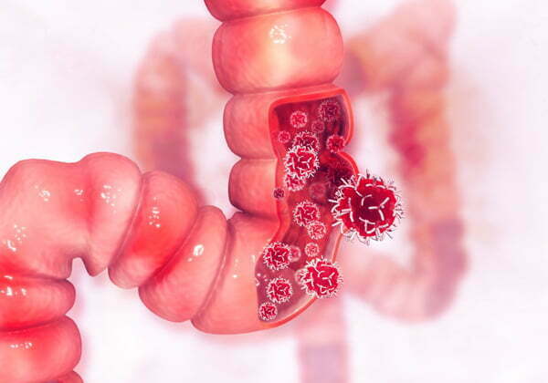
Think of it this way: the pathologist is a medical detective. They've taken a close look at your tissue sample under a microscope, and this report is their final case file, explaining exactly what they found. Its main purpose is to give you a definitive diagnosis and arm your care team with the details they need to map out your treatment.
Your First Look at a Complex Medical Document
Let's be honest, nobody expects you to understand this document perfectly on the first read-through. The goal here is to get familiar with the layout and the type of information it contains so you can have a more productive conversation with your doctor.
The report is structured logically, starting with general information about you and the tissue sample, then diving into the nitty-gritty details of what the pathologist observed.
What Does the Report Tell Us?
At its heart, the pathology report answers a few fundamental questions that will guide every decision that comes next. It’s much more than a simple "yes" or "no" on a diagnosis; it's the blueprint for your treatment.
- Is it cancer? This is the big one. The report will clearly state if the cells are benign (non-cancerous) or malignant (cancerous).
- What kind of cancer is it? If it is malignant, the report will specify the exact type, like adenocarcinoma or squamous cell carcinoma. The type matters—a lot.
- How aggressive does it look? You'll see a tumor grade, which is the pathologist's assessment of how abnormal the cells look. This helps predict how quickly the cancer might grow and spread.
- Where exactly is it? The report pinpoints the primary location and measures the size of any tumor found.
It’s a global challenge to make these reports easier to understand. That’s why organizations like the College of American Pathologists (CAP) have developed guidelines to standardize the information. This ensures that no matter where you are, your report contains the critical details—like tumor grade and staging—that directly influence treatment.
Key Takeaway: Your pathology report is not just a diagnosis—it's a comprehensive guide for your medical team. Each section provides a piece of the puzzle they use to build your personalized treatment plan.
Getting comfortable with the structure is the first step. It turns an overwhelming document into a powerful tool, helping you ask smarter questions and play an active role in your own care.
Before we dive into each section one by one, here's a quick overview of what to expect. Think of this table as your road map to the report.
Key Sections of a Pathology Report at a Glance
This table breaks down the main parts of a typical report, so you know what kind of information to look for in each one.
Having this high-level view can make the deep dive into the specifics feel much more manageable. Now, let's break down what each of these sections really means.
Breaking Down the Core Components of Your Report
Think of your pathology report as a story, with each section revealing a new chapter of your diagnosis. At first glance, it can feel like a dense, foreign language. But once you understand the structure, you can start to follow the narrative from the big picture down to the tiniest cellular details.
It all starts with the basics: your name, date of birth, and a unique medical record number. You'll also see your doctor's information and details about the specimen—the official name for the tissue sample taken from your biopsy or surgery. This part is all about triple-checking that the report belongs to you and the sample is correctly identified. It's a critical quality control step.
The First Look: Gross Description
Long before any high-powered microscopes come into play, the pathologist simply looks at the tissue sample. This is called the "gross examination," and the findings are logged in the Gross Description section. It’s what the pathologist can see, measure, and feel with their own eyes and hands.
This isn't a minor detail; it’s a crucial first assessment. The pathologist meticulously records things like:
- Size and Weight: How big is the tissue sample? The dimensions, usually in centimeters, are a key factor in staging a tumor.
- Color and Texture: Is the tissue firm or soft? Tan, white, or mottled? These physical characteristics can provide early clues.
- Ink and Location: Surgeons often use special colored inks to mark the edges of the tissue they remove. The pathologist notes these colors, which acts like a map to orient the sample and understand exactly where it came from in your body.
Essentially, the gross description is the pathologist drawing a map of the landscape before they zoom in. It sets the stage for everything that follows.
The entire diagnostic process is a journey, starting with that physical tissue sample and ending with a detailed report that guides your treatment.
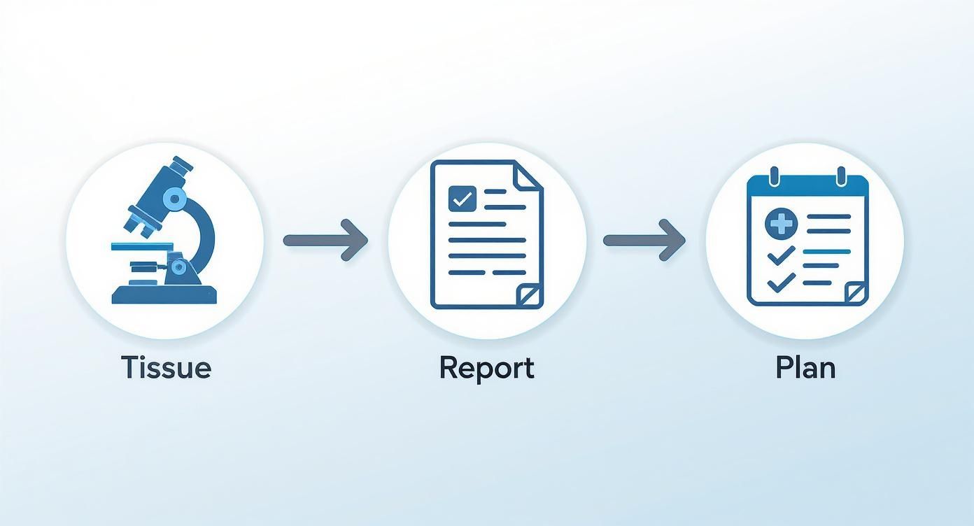
This workflow shows exactly how a piece of tissue is transformed into the critical information your care team uses to build a personalized strategy for you.
The Heart of the Report: Microscopic Description
Now for the main event. The pathologist takes incredibly thin slices of the tissue, stains them with special dyes to highlight different cellular structures, and examines them under a microscope. This is where the real detective work begins, and it's all captured in the Microscopic Description. Be prepared; this is often the longest and most technical part of the report.
Here, the pathologist is looking at the cells themselves, trying to answer the most fundamental questions about what’s going on at a cellular level.
Key Insight: The microscopic description is the raw evidence backing up the final diagnosis. It’s the "why" behind the conclusion, detailing how the cells look, how they're arranged, and how they're behaving.
You'll run into a lot of jargon here, but each term describes a specific feature. For example, cellular morphology just means the shape and structure of the cells. Do they look uniform and orderly like healthy cells, or are they misshapen and disorganized?
Another term you'll almost certainly see is differentiation. This describes how much the cancer cells still look like the normal, healthy cells they came from.
- Well-differentiated cells look a lot like normal tissue. This is generally good news, as they tend to be less aggressive.
- Poorly differentiated or undifferentiated cells look very abnormal and chaotic. These often grow and spread more quickly.
This section gives your oncologist a portrait of the cancer's "personality." It’s not just about knowing if cancer is present, but understanding how it's likely to act. This detailed picture is absolutely vital for predicting its behavior and figuring out which treatments will hit it hardest.
Making Sense of the Diagnosis: The "Bottom Line" of Your Report
After all the technical descriptions of tissues and cells, you finally get to the heart of the matter: the Final Diagnosis. Think of this as the pathologist's definitive conclusion. It’s the summary of everything they've observed and the answer to the big question: "What did you find?"
This section contains the most important terms you'll need to understand, as they truly set the stage for everything that comes next.
Benign vs. Malignant: The Most Critical Distinction
The first and most fundamental piece of information is whether the finding is benign or malignant. These two words carry a lot of weight and are the primary fork in the road for your medical journey.
- Benign: This is the good news you hope for. It means non-cancerous. A benign tumor or growth won't spread to other parts of your body. While it might still need to be removed depending on its size or location, it’s not typically life-threatening.
- Malignant: This is the medical term for cancerous. Malignant cells are unpredictable—they can grow without stopping, invade the tissues around them, and potentially travel (metastasize) to other areas of the body.
You might also come across terms like atypical or indeterminate. This is the gray area. It means the cells look unusual under the microscope, but they aren't clearly cancerous. This finding often leads to a "watch and wait" approach, more frequent monitoring, or sometimes additional testing to get a clearer answer.
A Note on Certainty: Pathologists are incredibly precise with their language. Phrases like 'consistent with,' 'suggestive of,' or 'features of' are chosen with great care to reflect their level of confidence. Interestingly, a 2021 study found that oncologists and patients often interpret these phrases with more certainty than the pathologist intended, which can affect treatment decisions. It's a good reminder of why asking follow-up questions is so important.
Getting into the Specifics of the Diagnosis
The diagnosis is rarely just one word. It will give a specific name to what was found, which is absolutely crucial for your treatment plan.
For instance, a report on a skin sample won't just say "cancer." It might specify "Basal cell carcinoma" or "Invasive melanoma." A breast biopsy might read "Invasive ductal carcinoma." Each of these names tells your doctor exactly what kind of cells have become cancerous and where they started.
Here’s how this plays out in real-world situations:
- Skin Biopsy: A diagnosis of "Melanoma in situ" is serious, but it means the cancerous cells are contained in the very top layer of the skin. This is a world away from "Invasive melanoma," where the cancer has already started to grow deeper into the tissue, requiring a much different treatment approach.
- Breast Biopsy: A report showing "Ductal Carcinoma In Situ (DCIS)" means abnormal cells are inside the milk ducts but haven't broken out. This has a very different prognosis and treatment plan compared to "Invasive Lobular Carcinoma," where cancer has already moved into the surrounding breast tissue.
The specific name of the cancer gives your oncologist a blueprint—it tells them how that particular cancer usually behaves and which treatments have the best track record against it.
Understanding Tumor Grade: How Aggressive Are the Cancer Cells?
Beyond just naming the cancer, the report will often assign it a tumor grade. This is not the same as the cancer stage. Grade tells you about the cancer’s personality—specifically, how aggressive it is.
The pathologist determines the grade by looking at how abnormal the cancer cells appear compared to healthy, normal cells. It’s usually on a scale of 1 to 3.
- Grade 1 (Low-Grade): The cells are well-differentiated. This is good news. It means they still look a lot like normal cells and tend to be slow-growing.
- Grade 2 (Intermediate-Grade): The cells are moderately differentiated. They look more abnormal and are expected to grow and spread at a moderate speed.
- Grade 3 (High-Grade): The cells are poorly differentiated or undifferentiated. They look very different from normal cells and are often disorganized. These cancers tend to grow and spread more quickly.
The tumor grade is a powerful piece of information that heavily influences your treatment plan. A high-grade tumor might call for more aggressive therapy, like chemotherapy, even if it was caught very early. This, along with other test results, helps your doctor predict your prognosis and the chances of the cancer returning.
Sometimes, the grade and other molecular details can point your team toward very specific treatments. You can learn more in our guide about how targeted therapy works by attacking cancer cells with particular characteristics.
Understanding Margins, Staging, and Advanced Test Results

Once you get past the main diagnosis, your pathology report dives into the details that truly shape your treatment plan. This is where we move from a general strategy to a highly personalized one.
Let's break down three of the most important parts: surgical margins, cancer staging, and the specialized tests that unlock modern therapies. These elements work together to give your oncologist a complete picture, guiding critical decisions about whether you might need more surgery, radiation, or specific medications.
What Are Surgical Margins?
When a surgeon removes a tumor, they don't just take the tumor itself. They also remove a small border of what looks like normal tissue surrounding it. This border is the surgical margin.
A pathologist then meticulously examines this rim of tissue under a microscope to check for any stray cancer cells. The result of this exam is one of the most critical pieces of information for determining if the surgery successfully got all the cancer out.
Think of it this way: the status of the margins tells your doctor if any cancer cells were likely left behind at the surgical site.
Negative Margins: This is the best-case scenario. It means no cancer cells were found at the outer edge of the tissue your surgeon removed. You might also see this called "clean" or "clear" margins.
Positive Margins: This means cancer cells were found right at the edge of the removed tissue. It’s a strong indicator that some cancer may still be in your body where the surgery was performed.
Close Margins: This is a bit of a gray area. It means cancer cells are very near the edge—often within a millimeter or two—but aren't touching it. For some cancers, this is treated with caution; for others, it may be acceptable.
The primary goal of any cancer surgery is to achieve negative margins. If your report comes back with positive or close margins, your oncologist will have a serious discussion with you about the next steps. This often involves either another surgery to remove more tissue or targeted radiation to destroy any cells left behind.
A Quick Guide to Surgical Margins
To help you understand what your margin status means for your treatment path, here’s a straightforward breakdown.
Understanding Surgical Margin Status
Knowing your margin status helps you understand if the first major step of your treatment—the surgery—accomplished its primary goal.
Decoding Cancer Stage with the TNM System
The stage of a cancer is a standardized way of describing its size and how far it has spread from its original location. It's important not to confuse this with the tumor grade, which describes how aggressive the cells look under a microscope.
Staging provides a universal language for doctors everywhere. The most common method they use is the TNM system.
Each letter gives your care team a key piece of information:
T (Tumor): This describes the size of the primary tumor. It's usually followed by a number from 1 to 4, with larger numbers meaning a bigger or more invasive tumor. You might see TX (tumor can't be assessed) or T0 (no evidence of a primary tumor).
N (Nodes): This tells you if the cancer has spread to nearby lymph nodes. N0 is great news—it means no cancer was found in the nodes. N1, N2, or N3 indicates that cancer cells have been found in an increasing number of lymph nodes.
M (Metastasis): This is the bottom line—has the cancer spread to distant parts of the body? M0 means no distant spread has been found. M1 means the cancer has metastasized to other organs, like the liver, lungs, or bones.
The pathologist combines these T, N, and M values to determine an overall stage, usually written as a Roman numeral from Stage I (very early) to Stage IV (advanced or metastatic).
For instance, a cancer classified as T1 N0 M0 is a small tumor that hasn't spread to the lymph nodes or anywhere else. This corresponds to a very early stage. This staging is fundamental to your care—an early-stage cancer might be handled with surgery alone, while an advanced stage will almost always require systemic treatments like chemotherapy.
Biomarkers, IHC, and a Look Inside Your Cancer
Modern cancer treatment goes far beyond just a tumor's size and location. Pathologists now run a whole battery of advanced tests to find specific molecular "fingerprints" on your cancer cells. These are known as biomarkers or tumor markers.
These tests hunt for specific proteins, gene mutations, or other substances that can be targeted by newer, smarter drugs. One of the most common techniques is Immunohistochemistry (IHC), which uses special antibodies to "stain" and light up certain proteins on cancer cells, making them visible to the pathologist.
A perfect example is in breast cancer, where a report will always list the status of three key biomarkers:
- Estrogen Receptor (ER)
- Progesterone Receptor (PR)
- HER2 (Human Epidermal Growth Factor Receptor 2)
If a tumor is ER-positive, it means estrogen is fueling its growth. That’s incredibly valuable information, as it opens the door to effective hormone-blocking therapies. Likewise, a HER2-positive cancer can be fought with drugs designed to attack that specific protein.
These tests are the bedrock of personalized medicine. By understanding the unique biology of your cancer, your oncologist can select treatments that are far more likely to work and often come with fewer side effects than traditional one-size-fits-all chemotherapy. To dig deeper into this, you can learn about the role of genomic testing in developing personalized treatment plans and see how it’s changing the future of cancer care.
Getting the Most Out of Your Doctor’s Visit

Knowing what the sections of your report mean is one thing, but the real understanding comes from talking it through with your doctor. This conversation is where the technical jargon on the page turns into a clear plan for you. Coming in prepared can make a huge difference.
Before your appointment, spend a little time with the report. Grab a highlighter or a pen and mark any words or phrases that don't make sense. Don't stress about what they mean just yet—the point is simply to flag what you need to ask about.
This simple act transforms a meeting that could feel overwhelming into a productive, focused conversation. It helps you become an active partner in your care, not just someone listening to a stream of information.
How to Ask Questions That Get Clear Answers
The way you frame a question often dictates the kind of answer you’ll get. If you ask vague questions, you'll likely get vague answers. The key is to be specific and connect the findings in the report directly to your situation and what comes next.
Think of it as getting to the "so what?" behind every piece of data. Here are a few examples of how to do that:
- For the Diagnosis: Instead of "What's my diagnosis?" try, "Can you explain what 'invasive ductal carcinoma' means in simple terms?" or "You mentioned the tumor is Grade 3. What does that tell us about how it might act?"
- For the Results: "The report says my margins are 'close.' What are the chances any cancer cells were left, and what would we do about that?"
- For Next Steps: "How do these biomarker results—the ER/PR and HER2 status—shape the treatment plan you're thinking of?" or "Looking at this whole report, what’s the single most important thing we need to do next?"
Jotting down your questions beforehand is a great strategy. It ensures you won’t forget anything in the moment and shows your doctor you’re invested and ready to work together.
Organizing Your Questions by Report Section
To keep the conversation on track, it can be helpful to group your questions by the main parts of the report. This creates a logical flow and makes sure you don't miss anything.
1. Getting Clear on the Diagnosis
- What is the specific type of cancer I have?
- What does the tumor grade mean for my prognosis?
- Is there anything in the diagnosis that’s still uncertain or needs more tests?
2. Understanding the Big Picture (Staging)
- Can you walk me through what the TNM staging in my report means for my treatment?
- My margins were positive/close. What's our game plan for that?
- Did the report show any spread to my lymph nodes or anywhere else?
3. Talking About Treatment
- How do my biomarker results guide our treatment options?
- Based on this report, am I a candidate for things like targeted therapy or immunotherapy?
- What are the main goals of the treatment you’re recommending?
An Empowering Tip: Always ask for a copy of your pathology report to keep. It's your medical information, and you have a right to it. Having it lets you review things later and prepare for other appointments down the road.
It's also interesting to know that technology is changing how this data is used. Researchers are now using machine learning and natural language processing (NLP) to analyze thousands of pathology reports at once, helping them spot patterns that could lead to new, personalized treatments. You can read more about how AI is making an impact in cancer research and making these reports even more powerful.
For an even more comprehensive list, take a look at our guide on the most important questions to ask your oncologist, which dives deeper into these crucial conversations.
Common Questions About Your Pathology Report
Even after going through a report piece by piece, it's completely normal to feel a bit overwhelmed when you have your own in front of you. These documents are packed with technical language, and let's be honest, the stakes are high. Let's walk through some of the questions I hear most often from patients to help you find your footing.
The first hurdle for almost everyone is the terminology. You hit a wall of words you've never seen before, and it can be discouraging. Don't let it be.
What Should I Do if I See a Word or Phrase I Don't Understand?
Your first instinct might be to pull out your phone and start searching. While that can give you a starting point, be careful. The internet doesn't know your specific situation, and medical context is everything.
A better approach is to simply jot down the exact word or phrase. If you want to do some initial research, stick to trusted sources like the National Cancer Institute for a reliable, general definition. But the most important step is to bring your list of questions to your oncologist.
A generic definition from a website can't tell you what a finding means for you. Your doctor can.
A pathologist might use a very specific term to describe a subtle feature they saw under the microscope. Your doctor is the one who can translate that observation into a clear "so what does this mean for me?" answer, connecting the report's findings directly to your treatment plan.
Can I Get a Second Opinion on My Pathology Report?
Yes, and you absolutely should if you feel you need one. Getting a second set of eyes on the pathology is a standard part of the process, especially with a serious diagnosis. It’s your right as a patient, and I can assure you, your care team will support you.
The process is straightforward. Your doctor’s office will help coordinate sending your original tissue slides to a pathologist at another medical center. That pathologist will then perform their own independent review and write a separate report.
This serves a couple of valuable purposes:
- Confirmation: More often than not, it confirms the original diagnosis, which can provide tremendous peace of mind.
- New Insights: Sometimes, a second review might offer a slightly different interpretation or highlight nuances that could open up different treatment possibilities.
Why Is There a Comment or Addendum Section?
I like to think of the Comment or Addendum section as a direct memo from the pathologist to your oncologist. It’s where they add important context that doesn't quite fit into the standardized fields.
For instance, the pathologist might use this space to explain why a diagnosis was particularly tricky or to suggest that another specific test might be helpful. An addendum is especially common; you’ll often see one added days or even weeks later once results from more complex tests—like IHC or molecular studies—are finalized. Pay close attention to this section, as it often contains critical information that can refine or even change the initial plan.
At Hirschfeld Oncology, we believe that understanding your diagnosis is the first step toward feeling empowered in your treatment journey. If you have questions about your pathology report or are seeking advanced care options, we are here to provide clarity and hope. Learn more and explore your options by visiting us at https://honcology.com/blog.
.png)
.png)