.png)


The core difference between a CT scan and an MRI really boils down to how they see inside your body and what they’re best at looking for. A CT scan uses X-rays to create incredibly fast and detailed pictures, making it a go-to for examining bones and other dense structures, especially in urgent situations. An MRI, on the other hand, uses powerful magnets and radio waves to get an exceptionally clear view of soft tissues like muscles, nerves, and the brain—all without using any radiation.
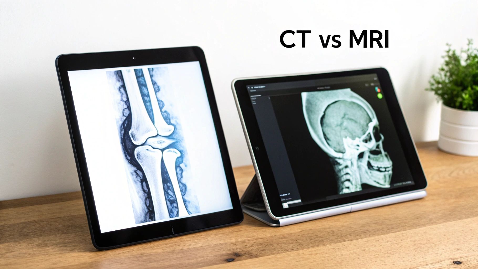
Understanding CT Scan vs MRI at a Glance
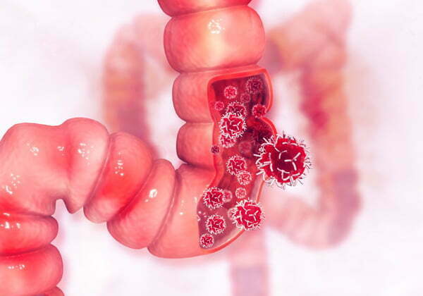
When you're navigating a cancer diagnosis, getting a handle on the imaging tests your doctor orders can make you feel more in control of your care. Both CT (Computed Tomography) and MRI (Magnetic Resonance Imaging) give us a detailed look inside the body, but they aren't interchangeable tools. Your oncologist will choose one over the other, and sometimes both, depending entirely on the specific question they need answered about your diagnosis or treatment.
Think of a CT scan as the workhorse of medical imaging, especially for initial cancer staging. It’s essentially a sophisticated, 3D X-ray machine that captures cross-sectional "slices" of your body in moments. Its speed is a huge advantage, giving us a quick yet thorough overview of the chest, abdomen, and pelvis to find tumors or check if cancer has spread.
An MRI is more like a high-definition specialist. It truly shines when we need to see subtle differences between healthy tissue and cancerous tissue. This makes it indispensable for looking at:
- Brain and spinal cord tumors
- Soft tissue sarcomas that arise in muscle or fat
- Certain cancers in the liver and pelvic region
Because it provides such incredible soft-tissue detail, an MRI is often the top choice for precise surgical planning and for tracking how well a treatment is working for specific cancers. And while a CT is great for spotting cancer that has spread to the bone, other specialized tests can also play a role. You can learn more about how a bone scan for prostate cancer fits into the bigger picture alongside these other scans.
The simplest way to think about it is this: A CT scan gives us a rapid, wide-ranging map of your body, while an MRI provides a focused, deep-dive investigation into specific areas.
To help put the core distinctions side-by-side, this table breaks down how the two technologies compare.
Quick Comparison CT Scan vs MRI
This summary table highlights the main differences between CT and MRI scans from a patient's perspective, covering the technology, speed, and typical uses in an oncology setting.
Ultimately, both scans are powerful diagnostic tools. The choice always comes down to getting the clearest possible picture to guide your specific cancer care.
How CT and MRI Scans Actually Work
To really grasp the difference between a CT scan and an MRI, you have to look at how each machine sees inside your body. They both produce incredibly detailed internal images, but their methods couldn't be more different. This fundamental distinction—one using radiation, the other using magnets—is what determines which scan your doctor will order.
The CT Scan: A Sophisticated X-ray
Think of a Computed Tomography (CT) scan as a super-powered, 3D X-ray. Instead of a single flat picture, a CT scanner sends a narrow beam of X-rays that rotates all the way around you. It captures hundreds, sometimes thousands, of images from different angles. A computer then acts like a master puzzle-solver, stacking these thin "slices" together to create a comprehensive cross-sectional image of your bones, organs, and tissues.
Since it’s based on X-ray technology, a CT scan is fantastic at seeing differences in tissue density. It can easily tell bone apart from muscle, or an organ from the air in your lungs. This is precisely why it’s the workhorse for quickly finding tumors, checking for internal bleeding, or getting a clear picture of the chest and abdomen.
The MRI: Harnessing Magnets and Water
An MRI (Magnetic Resonance Imaging) machine is in a completely different league. It uses zero ionizing radiation. Instead, its power comes from a massive magnet and radio waves. It’s a fascinating bit of physics, really. Your body is about 60% water, and the hydrogen atoms in that water act like tiny, spinning magnets.
When you slide into an MRI scanner, its incredibly strong magnetic field forces all those tiny hydrogen "magnets" to line up in the same direction. Then, a pulse of radio waves knocks them out of alignment. Once the pulse stops, the atoms snap back into place, releasing a faint energy signal that the scanner picks up.
Different tissues have different water content, so their hydrogen atoms realign at different speeds. The MRI’s computer translates these subtle timing differences into a remarkably detailed image, creating unparalleled contrast between different types of soft tissue.
Here's the bottom line: CT scans use X-rays to see density, making them great for a quick, clear look at bones and solid organs. An MRI, on the other hand, uses magnets to map out the water in your body, giving an amazingly detailed picture of soft tissues like the brain, nerves, and muscles.
A Tale of Two Technologies
This core difference—X-rays versus magnetic fields—is what drives every decision about which scan is right for you. It dictates what each machine is good at seeing and informs their safety profiles.
- CT Scan Technology: Uses a rotating X-ray tube. It builds images based on how different tissues absorb the X-rays, making it a master at showing structural details in dense materials.
- MRI Technology: Uses a powerful magnetic field and radio waves. It creates images based on the behavior of water molecules, allowing it to highlight subtle, nuanced differences in soft tissue.
The ionizing radiation in a CT scan is a factor, albeit a small one. While the dose for a single scan is low and considered very safe, your doctors always track cumulative exposure, especially if you need imaging often. This is a key reason why an MRI, which is radiation-free, is often the preferred choice for children or for long-term monitoring of certain conditions. This is the kind of practical consideration that guides your oncologist's choice every single time.
Choosing the Right Scan for Cancer Diagnosis
When you're dealing with a potential cancer diagnosis, the choice between imaging scans isn't about which one is "better." It's about which one will give your oncologist the clearest answers to very specific questions. The decision really boils down to the suspected cancer type, its location in the body, and the exact level of detail needed to build an accurate and effective treatment plan.
Think of it this way: a CT scan is like creating a quick, detailed anatomical map of a large part of your body. Its speed and fantastic ability to show bone, air, and solid organs make it the perfect starting point for getting a broad overview of the chest, abdomen, or pelvis.
An MRI, on the other hand, is more like a deep-dive investigation into a specific area. It truly shines when it comes to telling different kinds of soft tissues apart, often spotting subtle problems a CT scan might overlook. That high-contrast detail is priceless for looking at complex areas where absolute precision is a must.
When a CT Scan is the Go-To Tool
In oncology, getting information quickly across a wide area is often the first priority, especially when we're trying to figure out the stage of a cancer. A CT scan takes just minutes but gives us a huge amount of information about a tumor's size, its exact spot, and whether it has spread to nearby lymph nodes or other organs.
This makes it our first choice in several key situations:
- Lung Cancer: CT scans give us incredibly sharp pictures of the lungs, letting us see even tiny nodules and check if any nearby structures are involved.
- Abdominal Cancers: For cancers in the liver, pancreas, or kidneys, a CT provides a crystal-clear map of the organs and the blood vessels around them.
- Lymphoma: We rely on CT scans to map out the extent of swollen lymph nodes all through the body, which is a critical part of staging lymphoma.
A CT is also the workhorse for quickly checking if cancer has spread, or metastasized, to the bones. Because it’s so good at picking up changes in bone density, it’s a reliable first step for assessing skeletal involvement. For some pelvic cancers, a CT gives us vital information on tumor size and location. You can read more about how a CT scan is used to detect ovarian cancer and what it reveals in our detailed guide.
This simple decision tree helps visualize the core technology behind each scan—one uses X-rays, the other uses magnets.
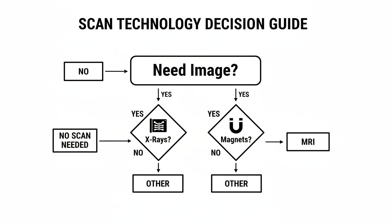
The flowchart breaks it down: if we need imaging based on X-rays, that points to a CT. If we need to use magnets for the picture, that's an MRI.
When MRI Offers Superior Insight
While the CT gives us the big picture, the MRI delivers the fine-grained detail we need for certain high-stakes diagnoses. Its ability to create contrast between different soft tissues is simply unmatched, making it the clear winner when the tiniest differences can change everything.
We almost always turn to an MRI for:
- Brain Tumors: An MRI can clearly separate tumor tissue from healthy brain and the surrounding swelling (edema). This is absolutely essential for both diagnosis and planning surgery.
- Spinal Cord Issues: If there's any worry that a tumor is pushing on or growing into the spinal cord, an MRI is the only tool that can show this with the needed clarity.
- Soft Tissue Sarcomas: These are cancers that start in muscles, fat, or other connective tissues. An MRI is indispensable for defining the tumor's exact edges and seeing how it relates to nearby nerves and blood vessels.
- Rectal and Prostate Cancers: For staging these cancers, an MRI gives us precise detail on how far a tumor has grown through the wall of the organ.
We opt for an MRI when we need exquisite detail. This isn't just for getting the diagnosis right; it directly shapes the surgical plan and helps our radiation oncology colleagues target tumors with pinpoint accuracy, saving as much healthy tissue as possible.
A CT offers a rapid, wide-ranging survey; an MRI provides an in-depth, focused investigation. Understanding this key difference helps clarify why your doctor might choose one, the other, or both to build a complete diagnostic picture.
Using CT and MRI Scans Together
In many cancer journeys, CT and MRI aren't rivals—they're partners. We often use them together to get the most complete picture of what’s going on. For instance, a patient might have a CT scan of their abdomen that shows a suspicious spot on the liver.
To get a better handle on it, an MRI is often the next step. The MRI can help us figure out if that spot is a harmless cyst, a primary liver cancer, or a metastasis that has spread from another cancer, like colon cancer. This two-step process plays to the strengths of both technologies—the speed and scope of the CT and the detailed characterization of the MRI—to help us reach a confident diagnosis and map out your care.
A Look at Radiation, Contrast Dyes, and Your Safety
When we talk about the difference between a CT scan and an MRI, safety is a top concern for both patients and clinicians. The conversation usually boils down to two things: radiation and the special dyes, or "contrast agents," used to make images clearer. Getting a handle on these two elements makes it much easier to understand why your oncologist might choose one test over the other.
The biggest safety difference is straightforward: CT scans use radiation, and MRIs do not. A CT scanner is essentially a very sophisticated X-ray machine, taking hundreds of pictures from different angles to build a detailed 3D image. This, by its nature, involves a small and carefully managed dose of ionizing radiation.
On the other hand, an MRI is completely radiation-free. It generates incredibly detailed images using a powerful magnet and radio waves. This makes it an especially safe choice for anyone who needs regular scans over a long period.
Putting Radiation Exposure in Context
The word "radiation" can sound scary, but the dose from a medical scan needs context. A typical CT scan delivers about 2-10 millisieverts (mSv) of radiation. To put that in perspective, it’s about the same amount of natural background radiation you’d absorb over one to four years just living your life. This difference is a major factor in healthcare planning; for instance, while CTs are often a go-to for speed, MRI use in U.S. hospitals still grew from 16.2 million scans in 2016 to 17.7 million in 2017. You can dive deeper into medical imaging statistics and trends on market.us.
While the risk from a single scan is minimal, your oncologist is always thinking about your total lifetime exposure. It’s why we use CT scans thoughtfully and often lean on MRI for children or for tracking chronic conditions that demand frequent imaging.
The core safety takeaway is simple: MRI is a radiation-free imaging technique, making it a go-to choice for sensitive populations and long-term surveillance. A CT scan's low radiation dose is considered safe, but exposure is always minimized when possible.
Why Contrast Agents Are Used
To get the sharpest possible images, your doctor might order your scan "with contrast." This just means a special dye will be given to you before the scan, which helps highlight specific tissues, blood vessels, or abnormalities. It makes it much easier for the radiologist to spot and analyze tumors.
It's important to know that CT and MRI don't use the same kind of dye. Each has its own unique properties and safety profile.
- For CT Scans: The dye is iodine-based. You might get it through an IV or, in some cases, as a drink. The iodine is great at absorbing X-rays, so areas with a rich blood supply (like many tumors) light up on the final images.
- For MRI Scans: The dye is gadolinium-based (GBCA). This agent works by changing the magnetic field in the water molecules of your body, which dramatically improves the detail and quality of the MRI images.
Safety Checks for Contrast Dyes
Both types of contrast are widely used and considered very safe for most people. However, your medical team will always go over your health history before giving you any contrast agent, just to be sure.
There are two main things they’ll be looking out for:
- Allergic Reactions: It's rare, but a small number of people can have an allergic reaction to the iodine-based dye used in CT scans. Reactions can be as mild as some itching or, very rarely, more serious. Be sure to tell your doctor if you’ve ever reacted to contrast dye or have a severe shellfish allergy.
- Kidney Function: Your kidneys are the organs that filter these dyes out of your system. If your kidney function is already compromised, using contrast requires a bit more thought. Your doctor will almost always check your kidney health with a quick blood test before ordering a scan with contrast.
In the end, the choice to use a contrast agent comes down to a simple risk-versus-benefit calculation. For most situations in oncology, the incredible diagnostic detail it provides is well worth the very small potential risk, as it allows your doctor to make the most accurate diagnosis and build the best possible treatment plan for you.
Preparing for Your Scan and What to Expect
Walking into an imaging appointment can be nerve-wracking, but knowing what’s coming can make all the difference. While the technology behind a CT scan and an MRI is vastly different, so is the patient experience. The prep work is usually simple and is all about making sure we get the clearest pictures possible.
Before either scan, you’ll need to remove all metal objects like jewelry, glasses, or removable dental work. Depending on what we’re looking at, you might be asked to fast for a few hours or drink a contrast agent to make certain organs stand out. Your care team will give you specific instructions ahead of time.
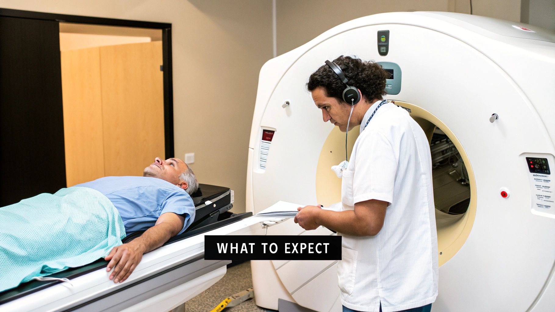
The CT Scan Experience
The first thing most people notice about a CT scan is how fast it is. The whole thing is often over in less than 15 minutes. You'll lie on a table that glides you through a large, open ring—it looks a lot like a giant donut. The machine is fairly quiet, just a soft whirring sound as it works.
Because the scan is so quick and the machine itself is so open, most people don't find it claustrophobic at all. This speed is exactly why CT is a workhorse in both routine cancer diagnostics and emergency situations.
Its efficiency and central role in medicine have driven major growth in the field. In fact, the global CT scanner market was valued at USD 7.28 billion in 2024 and is expected to hit USD 11.49 billion by 2032. If you're interested in the numbers, you can find more CT scanner market trends on Fortune Business Insights.
The MRI Scan Experience
An MRI is a different story. It’s a longer process, typically taking anywhere from 30 to 60 minutes, sometimes even longer for complex studies. You’ll lie on a table that slides into a longer, tunnel-like machine. The most important job you have during the scan is to stay perfectly still to avoid blurry images.
MRIs are also famous for being loud. You'll hear a lot of loud knocking, banging, and buzzing sounds from the magnets. Don't worry, you'll be given earplugs or headphones, and many centers will let you listen to music to help pass the time and block out the noise.
For patients who feel anxious in enclosed spaces, simple strategies can make a significant difference. Using an eye mask, practicing deep breathing exercises, or focusing on the music can help you relax and feel more in control during the scan.
Key Contraindications to Consider
This is where the differences between the two scans become critical for patient safety. Because MRI uses an incredibly powerful magnet, it’s not an option for everyone.
- MRI Contraindications: The biggest concerns are implanted metal devices. This includes older types of pacemakers, cochlear implants, certain aneurysm clips, and other electronic implants.
- CT Scan Contraindications: CT scans are generally safe for those with metal implants because there are no magnets involved. The main consideration is the small dose of radiation, which your medical team carefully calculates and manages.
It is absolutely vital that you give your medical team a full history, especially mentioning any implants, shrapnel, or even tattoos that might contain metallic ink. This information is key to keeping you safe and choosing the right scan for the job.
How to Discuss Your Imaging Plan with Your Doctor
Walking into a conversation with your oncologist armed with a basic understanding of CT and MRI scans can make a world of difference. It helps you become a true partner in your own care. The key thing to remember is that one scan isn't automatically better than the other; the right choice is always about asking the right clinical question.
Think of it this way: a CT scan gives us a quick, broad overview, almost like a geographical survey map. An MRI, on the other hand, is like zooming in on a specific neighborhood for a highly detailed street-level view, especially when it comes to soft tissues. Your doctor’s choice is a calculated one, based on your specific cancer, its location, and the exact information needed to make the best decision for you.
Preparing for Your Conversation
To get the most out of your time with your doctor, it’s a great idea to think through a few questions beforehand. This helps you cover all your bases and leave the appointment feeling clear and confident about what's next.
Here are a few questions you might want to ask:
- What are we hoping to learn from this scan? Is the goal to measure the tumor, see if it’s near other organs, or check if it has spread?
- Why is this specific scan the best choice for me at this stage? This helps you understand the reasoning for choosing a CT over an MRI (or vice versa) in your particular case.
- Will I need a contrast agent? If the answer is yes, you can follow up about what extra information it gives and mention any allergies or kidney concerns you might have.
- What will the experience be like? Asking about the duration, noise, and overall feel of the scan can do a lot to ease any anxiety.
Having a good back-and-forth with your oncologist is a cornerstone of great cancer care. When you feel comfortable asking questions, you gain a real understanding of each test, which helps you move forward with much more certainty and peace of mind.
Being an informed patient is one of the most powerful things you can be. For more ideas on how to approach these conversations, we've put together a list of questions to ask your oncologist that can help guide you. When you truly understand the plan, you become a collaborator in your own health journey.
Answering Your Questions
It's completely normal to have questions when you're navigating medical imaging. Understanding the difference between a CT scan and an MRI is a big part of feeling in control of your care. Let's walk through some of the things our patients ask us most often.
Can One Scan Just Replace the Other?
In short, no. CT and MRI scans are more like partners than competitors; they're not interchangeable. Each one gives us a unique piece of the puzzle. A CT scan is fantastic for getting a quick, wide-angle view of your anatomy, making it great for spotting a tumor's size and seeing if it has spread to organs or bones.
An MRI, on the other hand, zooms in on the fine details of soft tissues. So, a CT might be the first step to identify a suspicious mass, but an MRI often follows to get a much closer look. That extra detail helps us understand the character of the mass and determine whether it's cancerous or benign.
Is It Safe to Have Multiple CT Scans?
This is a really important question, and we hear it all the time. CT scans do use a low dose of ionizing radiation, and your care team is always tracking your total exposure over time. While the risk from any single scan is very small, we're careful when patients need regular imaging for follow-ups.
We always have to weigh the benefit of the information we get from the scan against the potential risk. If you need frequent monitoring, we'll often lean toward an MRI because it's completely radiation-free. Your oncologist’s job is to pick the safest and most effective tool for your long-term health.
Why Did My Doctor Order a Scan with Contrast?
When your doctor orders a scan "with contrast," it's to make specific parts of your body stand out more clearly. We use a special dye—usually iodine-based for CTs and gadolinium-based for MRIs—which is typically given through an IV. As this dye moves through your bloodstream, it lights up blood vessels and tumors.
Think of contrast agents as a spotlight for the radiologist. They illuminate the areas we need to see, which is absolutely critical for staging cancer accurately and planning precise treatments like surgery or radiation therapy.
Which Scan Is Better for Checking If My Treatment Is Working?
Both scans are essential for monitoring how well a treatment is working, and the best choice really comes down to the type of cancer and where it's located. For many solid tumors in the abdomen or chest, CT scans are the go-to for measuring any changes in tumor size. It's a straightforward way to see if a tumor is shrinking, staying the same, or growing.
However, for things like brain tumors or soft tissue sarcomas, an MRI is usually the preferred tool. Its incredibly detailed images can reveal subtle changes inside a tumor—signs that cancer cells are dying off even before the tumor itself starts to shrink.
At Hirschfeld Oncology, we know that an informed patient is an empowered patient. If you have more questions about your imaging plan or you're looking for a second opinion on advanced cancer treatments, we're here to offer clarity and compassionate care. To learn more about how we can help, please see the resources on our blog.
.png)
.png)