.png)


Early detection of stomach cancer often starts with a simple blood draw. By measuring tumor markers such as CEA, CA72-4, and CA19-9, clinicians can catch subtle changes long before symptoms appear. At the same time, ctDNA liquid biopsies are emerging as a powerful, noninvasive window into tumor behavior, guiding treatment choices at the earliest sign of trouble.
Primary Biomarkers Clinicians Track
Most doctors tailor their tests to each patient’s risk profile and treatment stage. The key assays include:
- CEA (Carcinoembryonic Antigen) to estimate overall tumor burden and watch for relapse, with 55% sensitivity in advanced cases.
- CA72-4 to sharpen detection of gastric tumors—often used alongside CEA to boost accuracy.
- CA19-9 to evaluate how well therapy is working, delivering 90% specificity in distinguishing cancer from benign conditions.
- ctDNA assays that isolate tiny DNA fragments shed by tumors, achieving higher sensitivity for spotting early-stage disease.

This image shows how a liquid biopsy pulls out circulating tumor DNA, sidestepping the need for a tissue needle.
Comparison Of Blood Tests For Stomach Cancer
Before diving into individual results, it helps to see how these tests stack up side by side.
Even within the same lab, these numbers can shift depending on tumor stage and testing methods. Combining markers often lifts overall detection rates and paints a clearer clinical picture.
Impact On Survival Rates
- Catching markers early can raise the 5-year survival rate in localized stomach cancer from 31% to 50%.
- ctDNA tracking may flag a recurrence 4–6 months before imaging scans show any change.
- Running multiple assays together pushes the positive predictive value up to 85%.
“Early intervention driven by biomarker trends can shift survival curves significantly,” note oncology teams in routine practice.
When To Order Specific Tests
- Patients with a strong family history should have baseline CEA and CA72-4 measured twice a year.
- Those on chemotherapy benefit from monthly ctDNA checks to spot minimal residual disease.
- After surgery, quarterly CA19-9 can catch a recurrence before symptoms arise.
Customizing the timing of each test helps clinicians stay one step ahead, adjusting therapy before disease progression gains momentum.
For a personalized testing plan or to discuss the role of blood assays in your care, reach out to Hirschfeld Oncology today.
Understanding How Blood Tests Detect Stomach Cancer
When cancer cells in the stomach grow or break down, they leave tiny clues in the bloodstream. Blood tests pick up on these signals—whether it’s a protein marker or a snippet of DNA.
Think of it like tracking footprints in the sand. A liquid biopsy “reads” those footprints to reveal where a tumor has been active.
How Tumor Markers Enter Blood
Cancer cells constantly turn over, releasing bits of themselves as they multiply or die. Those fragments include:
- Protein markers such as CEA, which often rise with greater tumor burden
- Carbohydrate markers like CA19-9 and CA72-4, now gaining traction in clinical practice
- Circulating tumor DNA (ctDNA) that carries the tumor’s unique genetic mutations
Once released, these markers drift through the bloodstream and can be captured with a simple blood draw. This noninvasive method stands in stark contrast to more intrusive procedures.
Benefits Of Liquid Biopsy
Liquid biopsies bring flexibility to cancer monitoring. You can draw blood at routine check-ups and spot changes long before a scan reveals a mass.
- Early Alerts: A shift in ctDNA levels can raise a flag weeks before symptoms or imaging changes
- Minimal Discomfort: No sedation or endoscope—just a quick blood draw
- Wide Access: Useful in clinics lacking advanced endoscopy units
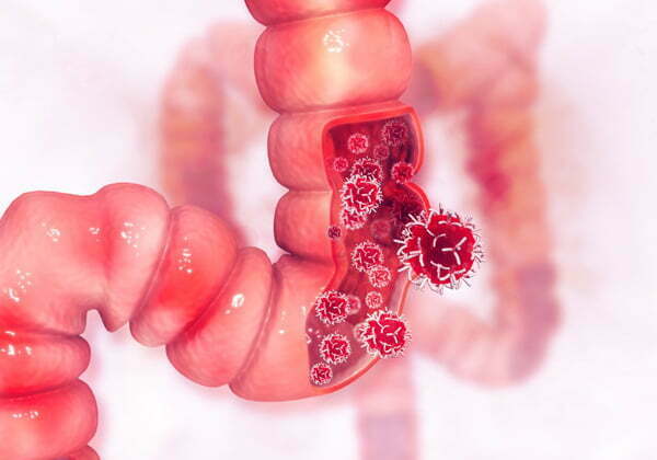
This image highlights how gastric tumors spread and why understanding anatomy is key to interpreting biomarker levels.
Comparing Blood Tests With Endoscopy
An endoscopy delivers a direct look at stomach lining under sedation. By comparison, a blood test is painless and can be done in minutes during an outpatient visit.
Stomach cancer (gastric cancer) remains the 5th most common cancer globally. In 2022, there were over 969,000 new diagnoses—about 4.8% of all cancers—and nearly 660,000 deaths, representing 6.8% of cancer mortality. For more details, see the stomach cancer statistics on WCRF.
“Combining blood tests with imaging can shift diagnosis earlier and improve outcomes.”
Validating New Biomarkers
Before any marker finds its way into routine care, researchers run multi-phase trials. They compare levels in healthy volunteers against those in patients with confirmed tumors. A typical validation process looks like this:
- Establish a cutoff point using healthy control data
- Calculate sensitivity and specificity in early versus late stages
- Reproduce results across different labs and diverse populations
When markers pass these hurdles, they can be woven into screening programs alongside scans—boosting early detection and helping decide when to biopsy.
Patients curious about blood-based screening should discuss personalized plans with Hirschfeld Oncology specialists. Early conversations can align monitoring schedules with individual risk profiles.
Integrating Tests Into Care Plans
Oncologists rarely rely on a single test. Instead, they blend blood-test trajectories with imaging results and patient symptoms. This layered method allows:
- Comparing rising marker curves to dashboard warning lights
- Synchronizing ctDNA checks with scheduled scans
- Using protein markers to gauge treatment response between imaging sessions
Work with your Hirschfeld Oncology care team to design a monitoring plan that fits your situation. Reach out to schedule a consultation and pick the right biomarkers for your needs.
Case Example Of Early Detection
A patient’s ctDNA levels began climbing subtly. That triggered an early imaging study, which found a sub-centimeter lesion. Because treatment started weeks sooner, the outlook improved dramatically.
Emerging markers—like miRNA signatures—are already in trials and may soon boost detection even further. Stay in touch with your oncologist for updates on the latest research and clinical studies.
Types Of Blood Tests For Stomach Cancer
Blood tests for stomach cancer detect specific molecules that tumors shed into the bloodstream. Think of these biomarkers as footprints in a forest—each one hints at a hidden source.
By comparing established assays with newer ctDNA panels, you get a clearer picture of how different markers reveal distinct traces. In practice, simple analogies make complex lab reports feel more approachable.
Key Established Biomarkers
For years, three protein markers have formed the backbone of gastric cancer blood testing.
- CEA (Carcinoembryonic Antigen) tracks overall tumor burden and flags potential relapse
- CA72-4 focuses on gastric tumor progression when paired with other assays
- CA19-9 gauges therapy effectiveness and highlights changes in treatment response
“Combining protein markers can lift overall detection rates and clarify clinical decisions.”
Example Case
A patient came in with vague upper-abdominal discomfort. Over three months, rising CEA levels prompted imaging that uncovered a small gastric lesion—removed before it could advance.
Emerging Ctdna Panels
Liquid biopsy taps into circulating tumor DNA (ctDNA) fragments, offering a genetic window into cancer activity. It’s like finding shredded pages from a suspect’s diary mixed into a pile of ordinary documents.
By tracking ctDNA over time, clinicians catch early recurrences or confirm whether a treatment is working.

This infographic shows how ctDNA and protein markers combine noninvasive sampling to pinpoint tumor signals. Blending these approaches raises early detection rates by capturing multiple “footprints.”
When To Choose Each Test
Selecting the right assay depends on risk factors and clinical goals.
Screening At-Risk Patients
- Start with baseline CEA and CA72-4 for those with a family history.
Confirming Diagnosis
- Add CA19-9 alongside imaging when symptoms or protein markers are elevated.
Monitoring Treatment Response
- Schedule monthly ctDNA panels to spot subtle shifts before they appear on scans.
- Variability in marker expression between patients
- Overlapping elevations in benign diseases
- No universally accepted cutoff values for some assays
- How often to repeat blood draws to track trends
- Integrating results with endoscopic or imaging schedules
- Genetic profiling options and potential trial enrollment
- CEA: up to 5 ng/mL is considered normal. Values above may hint at tumor activity.
- CA72-4: typically under 6 U/mL. A rise calls for further checks.
- CA19-9: you want to see under 37 U/mL. Higher levels can reflect inflammation or malignancy.
- Patients over 60 may have naturally elevated baselines
- A diet rich in processed meats can skew results
- Flare-ups of gastritis often create temporary spikes
- Review trending blood marker levels
- Match against reported symptoms
- Order imaging or biopsy if indicated
- Sharpens early-detection accuracy
- Cuts down unnecessary invasive procedures
- Guides personalized care plans
- Scheduling a CT or MRI in 4–8 weeks
- Repeating blood tests to monitor marker trends
- Planning an endoscopic biopsy if levels keep climbing
- Keep a log of symptoms and test dates
- Ask questions if marker levels shift unexpectedly
- Request reports that clearly list reference ranges
- Persistent upper abdominal discomfort lasting more than two weeks may warrant a CT scan or endoscopy.
- Sudden spikes in CEA or CA72-4 above a patient’s baseline often signal renewed tumor activity.
- Unexplained weight loss of more than 5% body mass or new-onset anemia are critical alerts.
- Compare persistent symptoms against biomarker trends.
- Schedule imaging (CT or MRI) when markers rise or discomfort worsens.
- Follow up with endoscopic biopsy to confirm any suspicious lesions.
- CEA Levels track tumor burden and recurrence risk. Even a small uptick can hint at new growth.
- ctDNA Fractions measure genetic fragments shed by cancer cells. A rise of just 0.5% often appears weeks before imaging detects changes.
- CA19-9 and CA72-4 reflect chemotherapy response. Drops of 20% or more frequently mean treatment is hitting its target.
- Reviewing marker trends against past results
- Repeating tests in 2–4 weeks to confirm any spikes
- Correlating blood data with imaging and symptoms
- Schedule blood draws at consistent intervals—often monthly
- Keep a diary of side effects; they can mimic marker shifts
- Ask for both absolute values and percentage changes for the clearest picture
- Monthly blood tests alternating with quarterly imaging
- Telehealth check-ins to review marker trends promptly
- Expanded genetic panels to catch emerging mutations early
- Month 1: Baseline markers
- Month 2: CA72-4 down 30%
- Month 3: ctDNA plateau triggers imaging
- Month 4: Scan shows response
- Combined assays can pick up up to 85% of cases.
- Inflammation or noncancerous conditions sometimes trigger false positives.
- Tiny tumors may not release enough protein to register, causing false negatives.
- Fast for 8–12 hours before your appointment.
- Stop biotin and similar supplements if your doctor recommends it.
- Let your provider know about any recent infections or new medications.
- Most insurance plans cover markers when accompanied by a valid diagnosis code.
- Newer circulating tumor DNA tests sometimes require extra approval.
- Medicare usually reimburses conventional assays.
- Repeat tests in 4–6 weeks to confirm any upward or downward trends.
- If markers continue to climb, your doctor might order a CT scan or MRI.
- At that point, an endoscopic biopsy often provides definitive answers.
You might be interested in learning more about the impact of genomic profiling on gastrointestinal cancer treatment.
Early detection driven by biomarker trends can shift survival outcomes substantially.
Real-World Insights
Most gastroenterology centers pair blood tests with imaging to get a full clinical picture.
In one case, a mid-stage patient on chemotherapy showed a drop in CA19-9 but stable ctDNA. That mismatch triggered an early scan and led to a timely change in therapy, extending progression-free survival.
Regular blood testing delivers near-real-time feedback on tumor behavior, reducing uncertainty and guiding treatment tweaks.
Diagnostic Limitations
Blood tests aren’t foolproof. Protein markers can spike due to inflammation or non-cancerous conditions. Small tumors in early stages might not shed enough markers, leading to false negatives.
Key challenges include:
A blood test should complement—not replace—a tissue biopsy. Clinicians rely on them to decide when to schedule imaging or direct sampling.
Tip
Always interpret blood test results alongside clinical symptoms and imaging findings.
Next Steps With Your Oncologist
After reviewing your blood test, discuss:
Scheduling a consultation at Hirschfeld Oncology can help you design a monitoring plan tailored to your risk and treatment phase.
Consultation And Support
Hirschfeld Oncology offers one-on-one sessions to break down your test results. Their team analyzes biomarker trends and maps out a clear follow-up schedule.
Patients value direct access for quick questions.
“Access to real-time blood test interpretations eased my anxiety,” says a recent patient.
Telehealth visits are available for those outside Brooklyn or with busy schedules.
Interpreting Blood Test Results
Reading blood work for gastric cancer is a bit like tuning into a car’s dashboard. You’re looking for signals in three key markers—CEA, CA72-4, and CA19-9—to see if anything is drifting off its normal path.
Imagine each marker as a traffic light: green within range, yellow when you need a closer look, red if it’s time to act. Here are the usual cutoffs:
Of course, these numbers can shift slightly between labs or if factors like age, diet, or a recent infection are at play. If a result hovers near the threshold, it often makes sense to retest in four to six weeks before drawing conclusions.
Interpreting marker trends in context of symptoms and imaging prevents premature alarm, says Dr. Hirschfeld.
Common Factors Affecting Results
Real life rarely follows a neat script. Smoking, obesity, and even colds can nudge these markers higher—no cancer required.
On the flip side, very small tumors might not release enough proteins or ctDNA to register, leading to false reassurance.
That’s why an isolated abnormal value usually triggers follow-up rather than an immediate treatment change. Physicians combine lab numbers with clinical signs, scans, and pathology to keep both overreaction and oversight at bay.
When the pattern suggests real concern, targeted imaging—like CT or PET scans—often comes next:
Integrating Tests With Clinical Context
Blood results tell part of the story. Symptoms such as persistent pain or unexplained weight loss fill in crucial details.
Together they help decide whether to book a scan or schedule an endoscopy. This approach:
When blood tests alone aren’t definitive, imaging and tissue sampling step in. A multidisciplinary team—pathologists, radiologists, oncologists—then pieces all the data together.
Accurate interpretation balances lab data with patient history to minimize both oversights and overtreatment.
Next Steps After Interpretation
If your markers raise a red flag, your oncologist at Hirschfeld Oncology will map out the follow-up plan. This often includes:
Before each appointment, you might fast or pause certain supplements that can skew results. Clear notes on your diet, current meds, and any new symptoms ensure your team sees the full picture.
From there, you’ll settle on a monitoring rhythm—often monthly blood draws alternating with imaging. Think of it as charting a stock portfolio: steady slopes may trigger early treatment adjustments before tumors grow too big.
Blood tests are one tool among many—including imaging and pathology—to keep watch. If anything looks out of place, reach out to Hirschfeld Oncology for a detailed review.
Clear interpretation of blood tests empowers you to partner actively in your care.
Stay curious. Stay prepared. And always feel free to ask.
When To Use Imaging And Biopsy
Blood tests offer vital hints about gastric cancer, but they don’t tell the whole story. Biomarker levels can shift from one lab to another or vary between patients. A “normal” result doesn’t rule out disease; false negatives can and do occur.
Recognizing Red Flags
For example, someone with mild indigestion and rising markers could need imaging—even if an endoscopy looked clear six months earlier. Timing this escalation properly balances noninvasive monitoring with precise tissue diagnosis.
Timing Diagnostic Decisions
According to SEER data, only 31% of stomach cancer cases are caught at a localized stage, where the five-year survival rate sits at 76.5%. Another 24% are regional (five-year survival 37.2%), and 36% reach distant spread (survival 7.5%). Early blood tests targeting key biomarkers might help shift diagnoses into those earlier, more treatable categories.
Diagnostic delays reduce options and outcomes; early imaging preserves therapeutic choices.
Survival Rate By Stage For Stomach Cancer
Below is a concise look at how stage at diagnosis drives long-term survival.
This table underscores why moving from blood tests to imaging or biopsy at the first sign of trouble can be life-changing.
Next Steps After Testing
Imaging pinpoints targets that blood tests only hint at, paving the way for focused biopsies and accurate staging. If questions linger after initial labs, reach out to Hirschfeld Oncology for a tailored diagnostic plan. Their team integrates blood tests, scans, and pathology to craft follow-up intervals that fit your risk profile.
Early action on red flags not only boosts treatment options but also eases patient anxiety. Take, for instance, Case Study A: a 58-year-old with a sudden CEA jump post mild indigestion underwent a prompt CT. A tiny lesion was spotted, biopsy confirmed early gastric cancer, and a localized resection led to an excellent prognosis.
For a personalized diagnostic pathway, call Hirschfeld Oncology at 718-XYZ-1234 or visit Hirschfeld Oncology Blog. Booking imaging and biopsy consultations now can make all the difference.
Monitoring Disease And Guiding Treatment Decisions

Serial blood tests after a stomach cancer diagnosis offer a live update on tumor activity. Rather than waiting for scans, you watch markers climb or fall in real time.
Think of these results like weather maps for your treatment journey. Shifts in CEA or ctDNA resemble fronts moving in—signaling storms of progression or calm remissions.
Key Biomarkers To Watch
“Regular ctDNA tracking flagged a relapse four months before scans did,” says Dr. Hirschfeld.
Interpreting Trend Patterns
Oncologists read these marker curves like a dashboard. A steady climb in ctDNA might trigger a therapy switch. A plateau or dip can support continuing the current plan.
Next steps usually include:
Short-lived fluctuations sometimes stem from infections or lab variability. That’s why sticking with the same lab and methods is crucial.
Subtle curves can act as early warning lights. Always bring unexpected trends to your oncologist’s attention.
Integrating With Treatment Decisions
Blood markers rarely stand alone. Your care team weaves these numbers with scan reports, pathology findings, and how you feel day to day.
Read more about gastric cancer treatment options in our guide on Hirschfeld Oncology
Practical tips:
Consider this real-world case: ctDNA rose by 0.8% while CA19-9 fell by 15%. That mixed signal prompted an early PET scan, which found a small lesion. A timely therapy switch then drove markers back to zero.
Next Steps In Your Care
Work with your medical team to build a dynamic monitoring plan. Options might include:
“Dynamic monitoring turns static snapshots into a living report,” notes a Hirschfeld Oncology specialist.
Staying proactive lets you use blood tests as an early alert system. Small adjustments based on marker shifts often translate into better outcomes and more treatment choices down the line.
Case Study Insight
In one Hirschfeld Oncology case, a 62-year-old patient experienced a 30% drop in CA72-4 after two chemotherapy cycles, while ctDNA levels plateaued over four weeks.
The team maintained the same regimen. A follow-up PET scan then revealed tumor shrinkage that aligned perfectly with those blood marker trends.
Timeline Of Events
Structured monitoring can catch relapses early and expand treatment options. Contact Hirschfeld Oncology to tailor your monitoring plan today for better outcomes and confidence in care.
Common Questions About Blood Test For Stomach Cancer
Blood tests often raise more questions than answers, especially when you’re worried about stomach cancer. This section walks you through the most pressing concerns, step by step, so you know exactly what to expect.
Test Reliability And Limitations
No lab report is flawless—but by combining multiple markers, we narrow down uncertainty. Think of it like overlapping safety nets: each marker fills in the gaps left by another.
Preparation Tips For More Accurate Results
A few simple habits can make your numbers more trustworthy. Treat your blood draw like tuning a musical instrument: little adjustments go a long way.
Insurance Coverage And Cost Considerations
Understanding costs and coverage can feel like decoding a secret language. Here’s a quick breakdown:
“Discuss abnormal results early to avoid delays in imaging or biopsy,” advises Dr. Hirschfeld.
Don’t let billing questions stand in the way of timely testing.
Discussing Abnormal Results And Next Steps
A single elevated marker isn’t a verdict—it’s a signal to dig deeper. Tracking changes over time gives your care team a dynamic view of what’s happening inside.
Our approach always centers on personal guidance. If you’re wondering how these blood tests shape treatment, check out our detailed overview of chemotherapy adjustments. You might find our guide on chemotherapy for stomach cancer in our article on Hirschfeld Oncology here.
Ready to take the next step? Reach out to Hirschfeld Oncology at https://honcology.com/blog for tailored advice and ongoing support.
.png)
.png)