.png)


Fibroids almost never turn into cancer, with the chance of a fibroid being a cancer such as leiomyosarcoma commonly described as less than 1 in 1,000, or about 0.1%. A key concern is that a rare leiomyosarcoma can look like a fibroid before surgery, so the question is often about misclassification, not transformation.
That distinction matters because a patient can hear “fibroid” and assume the story is over, while the clinical question is still open. Can fibroids turn into cancer? The short answer is no in the usual sense, but the longer answer is the one that protects patients, because a growth that seems routine can sometimes need a closer look.
The Short Answer Most Patients Need
Fibroids almost never turn into cancer. What worries doctors more is a rare uterine leiomyosarcoma being mistaken for a fibroid before surgery.
That's why this topic deserves more than a quick reassurance. A fibroid is usually a benign muscle tumor in the uterus, and the cancer concern comes from a different tumor that can share symptoms and imaging features. If you've been told you have a fibroid, the next question is not “How do fibroids mutate?” It's “Does this mass fit the usual fibroid pattern, or does it need a specialist review?”
A calm answer still has limits. Most fibroids stay noncancerous, but a growing mass, new bleeding after menopause, or pain that doesn't match your usual pattern deserves attention. Imaging can help, but it cannot rule out sarcoma in every case, which is why doctors sometimes need tissue diagnosis after surgery or a more specialized workup.
Practical rule: reassurance is strongest when symptoms are stable, age and menopausal status fit the usual fibroid story, and imaging looks typical. The more the picture changes, the more the evaluation should change with it.
This article walks through the biology in plain language, the accurate risk numbers, the warning signs that matter, and the point where a second opinion stops being optional and starts being smart. It also separates what online articles often blur together, the difference between a fibroid becoming cancer and a cancer being present from the start.
What Fibroids and Leiomyosarcoma Actually Are
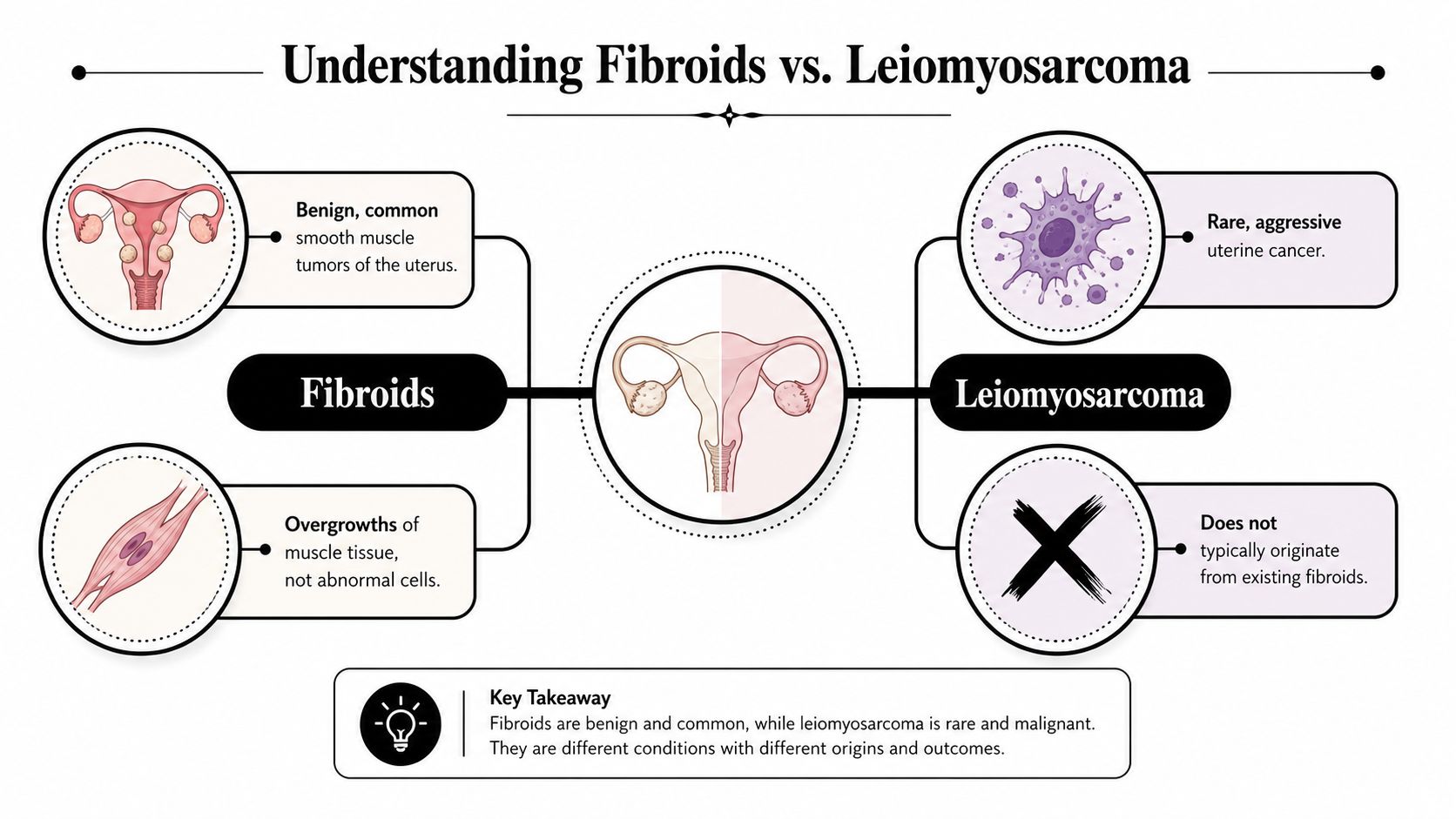
A fibroid is a benign smooth-muscle tumor of the uterus. Think of it as a knot of muscle cells that grew in the wrong pattern, but not as cells that have learned to behave like cancer. Mayo Clinic states that fibroids are not cancer and almost never become cancer, and Johns Hopkins Medicine says the same thing in plain clinical language. Mayo Clinic on uterine fibroids, Johns Hopkins Medicine on uterine fibroids
A useful analogy is a garden hedge versus poison ivy. A fibroid is an overgrown hedge, bulky and unwanted, but still made of the same kind of plant material as the rest of the yard. Cancer is poison ivy in the same yard, it may occupy the same space, but it behaves differently, spreads differently, and needs a different response.
Leiomyosarcoma is the dangerous counterpart here. It is a rare, aggressive uterine cancer that also comes from smooth muscle, which is why the two can be confused on scans or even during surgery. The key point is that sharing a tissue of origin does not make them biologically similar. Fibroids are not considered a precancerous lesion, and they are not the kind of growth that is expected to march toward malignancy over time.
Why the wording gets people tangled up
When someone says “cancer in a fibroid,” they may be using shorthand for a much more complicated situation. Sometimes the mass was never a fibroid at all, it was a sarcoma that looked fibroid-like from the beginning. That's why the exact wording matters so much in real patient conversations.
Plain-language translation: fibroids are benign muscle growths, leiomyosarcoma is a separate cancer, and the overlap is mainly about appearance, not biology.
The Actual Risk Numbers in Plain English
The number most patients can hold onto is less than 1 in 1,000, or under 0.1%, for a fibroid being cancerous. That figure is the best practical benchmark in the provided sources for the risk of a “cancerous fibroid.” Risk of cancer with fibroids
That does not mean every suspicious mass is harmless. The FDA revised risk estimates for unsuspected uterine sarcoma to 1 in 352 and uterine leiomyosarcoma to 1 in 498 in the specific setting of surgery for presumed fibroids. Those are not the same thing as the overall chance that a fibroid transforms into cancer. They describe a different clinical problem, the possibility that a mass thought to be a fibroid was a sarcoma all along. Fibroids and cancer risk discussion
Here's the plain-English way to read those numbers. The first number helps answer the fear of transformation. The other numbers help doctors think about surgical planning and diagnostic uncertainty when a mass is being removed.
| Clinical Scenario | Reported Risk | What It Actually Means |
|---|---|---|
| A fibroid becoming cancer | Less than 1 in 1,000 | Fibroids are overwhelmingly benign, and true malignant change is very uncommon |
| Unsuspected uterine sarcoma in surgery for presumed fibroids | 1 in 352 | A mass may have been misclassified before surgery |
| Uterine leiomyosarcoma in surgery for presumed fibroids | 1 in 498 | A rare cancer can resemble a fibroid clinically or on imaging |
| Uterine leiomyosarcoma in the U.S. overall | About 6 per 1 million women per year | The cancer itself is rare at the population level |
A diagnosis can still be scary even when the odds are low. That's why risk should be interpreted alongside age, menopausal status, symptom pattern, and imaging, not in isolation. If you want a clear primer on how to think about small and large odds without overreacting, this explanation of absolute risk versus relative risk is a helpful companion.
Why “Fibroid Cancer” Is Usually a Different Story
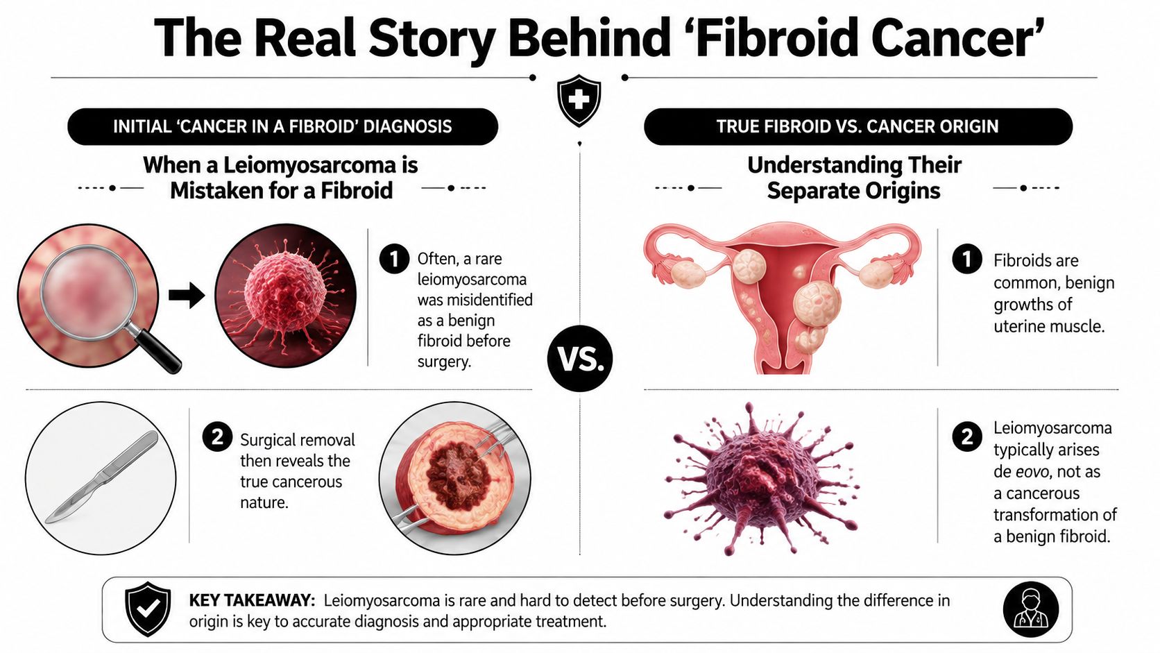
When a patient is told a fibroid “was cancer,” the more common explanation is that the cancer was already there and the mass looked like a fibroid before surgery. The clinical problem is usually misdiagnosed uterine sarcoma, not a benign fibroid that slowly changed into cancer later. Fibroids and cancer review
That distinction matters because it changes how doctors interpret imaging, choose a procedure, and read pathology. A benign fibroid and leiomyosarcoma can both present as a uterine mass, and ultrasound or MRI does not always separate them cleanly. The primary concern is often preoperative misclassification, where a suspicious growth is labeled a fibroid because it looks ordinary at first glance.
Why association is not the same as causation
A PubMed-indexed review reported more later cancer diagnoses among women treated surgically for fibroids, including cancers of the breast, uterus, ovary, kidney, thyroid, peritoneum, and retroperitoneum. That finding does not show that fibroids become those cancers. It shows an association in a treated group, which still needs careful interpretation and more study. Fibroids and cancer review
That is why a fibroid label should never end the discussion if the story feels off. The next question is whether the timing, symptoms, and imaging fit a routine benign pattern or whether the mass deserves a cancer-focused workup.
The mental model that helps
A fibroid is usually a benign growth that stays in one lane. A sarcoma is a different disease that can wear the same costume, which is why the diagnostic problem is so often about resemblance rather than transformation. That is also where pathology review matters, and why a clear guide to how to read a pathology report can help patients understand what was found.
In a Brooklyn oncology practice, that distinction often shapes the next step in the patient pathway. One team may review imaging, another may coordinate surgery or pathology, and a second opinion can help sort out whether the mass behaved like a fibroid or whether it needed a more careful cancer evaluation. Emerging research, including work around MED12, also adds to the picture by helping doctors understand why some tumors look like classic fibroids while others do not.
When patients understand that difference, they stop asking, “Did my fibroid become cancer?” and start asking the more useful question, “Does this mass need a cancer-focused evaluation?”
Warning Signs and When to Ask for More Tests
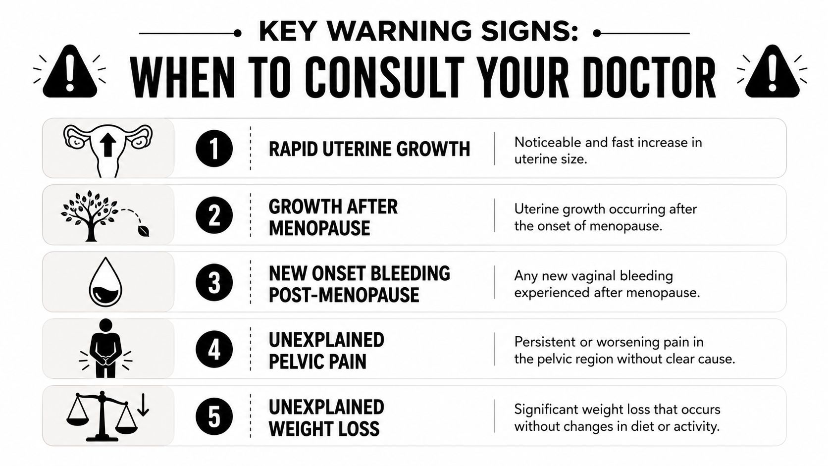
The red flags are less about fear and more about pattern recognition. A fibroid that has been quiet for years and then starts changing after menopause deserves a different level of attention than a stable, long-standing one. Johns Hopkins Medicine notes that imaging cannot reliably exclude sarcoma in every case, so the decision often comes down to how the story fits together, not one scan alone. Johns Hopkins Medicine on uterine fibroids
Signs that should prompt a call
- Rapid uterine growth: a uterus that seems to enlarge noticeably over a short period, especially if it's new for you.
- Growth after menopause: a mass that keeps getting bigger after periods have stopped.
- New bleeding after menopause: any vaginal bleeding that starts after menopause should be checked.
- Persistent pelvic pain: pain that doesn't settle, changes character, or keeps worsening.
- A mass that looks atypical on imaging: unusual borders, uneven internal appearance, or a pattern your clinician says doesn't look like a routine fibroid.
Ultrasound is often the first test, because it is accessible and can show the size, number, and location of fibroids. MRI can add detail when doctors need a better look at tissue pattern, blood flow, or whether the mass has features that feel off. If bleeding is part of the picture, tissue sampling or surgical pathology may be needed to get a definitive answer.
What usually happens next
The right next step depends on the whole picture, not just the word “fibroid.” If symptoms are mild and the imaging fits a typical benign pattern, careful observation may be reasonable. If the story is atypical, a gynecologic oncologist or other specialist may be the person who helps sort out whether more testing or surgery is needed.
For anyone trying to understand how scan findings become a pathology report, this guide on how to read a pathology report can make the language less intimidating.
Postmenopausal Bleeding and Other Subtle Red Flags
The gray zone is where patients most often feel dismissed. A little spotting around the menopausal transition can happen for benign reasons, but bleeding that starts after menopause needs a closer look because the normal hormone-driven explanation is no longer there. If you're trying to sort out whether a change is part of transition or something that needs evaluation, this discussion of perimenopause spotting between periods gives useful context.
The same logic applies to a fibroid that seems to change after menopause. Most fibroids are benign, but a new growth pattern after periods have stopped is one of the situations that makes doctors pause. It's not proof of cancer. It is a reason to stop assuming the old diagnosis still fits.
Recent reviews also point to a more technical future. Researchers are studying histopathology, cancer biomarkers, advanced imaging, and gene alterations such as MED12 to better separate benign fibroids from malignant smooth-muscle tumors before surgery. CEOG review on fibroids and sarcoma separation
Clinical reality: “uncertain” is not a weak diagnosis, it's a signal that the next step should be more expert, not more casual.
That matters because patients often hear, “It's probably fine,” when what they really need is, “It's probably benign, but we should check the features that make this one different.” Subtle red flags are the bridge between reassurance and caution. They're also the reason a specialist may choose a more careful imaging review, tissue confirmation, or surgical planning rather than relying on a single scan alone.
How a Second Opinion and Oncology Coordination Helps
A patient might walk into a second opinion visit with a scan disk, a pathology report, and a lot of worry. A good oncology consultation doesn't start by assuming the worst. It starts by asking what has already been seen, what has already been removed, and whether the current evidence really matches a benign fibroid story or needs a different lens.
If pathology is already available, the review focuses on the exact wording, because the details matter. If surgery hasn't happened yet, the discussion usually centers on whether imaging is convincing enough for observation, whether more testing is sensible, or whether surgery should be planned in a way that keeps a rare cancer from being spread unknowingly. If a sarcoma is confirmed, treatment planning becomes a different conversation, one that may include surgery, surveillance, and carefully selected systemic therapy.
A Brooklyn practice like Hirschfeld Oncology can fit into that pathway by coordinating with the OB/GYN, radiologist, surgeon, and primary oncologist rather than replacing them. That matters when decisions are nuanced and timing matters, because the patient benefits when expertise layers together instead of competing. For patients who've had confusing records or translated reports, resources on critical medical translation liability issues are a reminder that clear language can affect real care decisions.
If molecular testing enters the conversation, this overview of what molecular testing is and how it shapes personalized treatment can help explain why doctors may look beyond the basic diagnosis.
What to Do Next and What to Let Go Of
Let go of the idea that every fibroid is a ticking cancer risk. Fibroids are overwhelmingly benign, and the main challenge is usually making sure a rare sarcoma isn't being mistaken for one before surgery. What you should keep is a sharper question set, because context matters more than the label alone.
If you have new bleeding after menopause, a mass that seems to be changing, or pelvic pain that doesn't fit your usual pattern, call your clinician. Ask what the scan shows, whether the timing makes sense for a benign fibroid, and whether a gynecologic oncology review would add value. If the answer feels vague, a second opinion is reasonable.
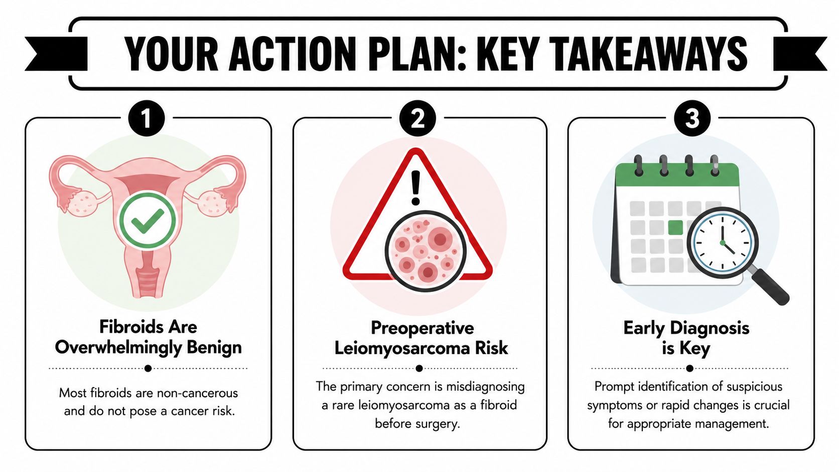
The simplest summary is this. Fibroids almost never turn into cancer, but suspicious changes deserve respect because the diagnostic problem is real. If you want a careful review of imaging, pathology, and next steps, visit Hirschfeld Oncology to learn how the team approaches complex cancer questions with patient-centered coordination and clear guidance.
.png)
.png)