.png)


A 7 tesla MRI can change what an oncologist sees, but only when the question is narrow enough for ultra-high-field imaging to matter. In a systematic review and meta-analysis, 7T MRI showed a 65% lesion-detection rate versus 22% for 1.5T or 3T MRI, a 33-percentage-point gap that can alter staging, trial eligibility, or whether a small lesion is even counted at all (Nature technical report). For patients with advanced cancer, that difference matters most when the target is tiny, the anatomy is complex, and the treatment decision hinges on details that a standard scan can miss.
Why 7 Tesla MRI Matters for Advanced Cancer Care
A cancer scan is only useful if it changes a decision. That's where 7 tesla MRI earns attention. In brain-heavy oncology questions, the difference between seeing a subtle lesion and missing it can change whether a patient is considered for surgery, radiation, a trial, or a more focused systemic plan. The strongest evidence for that advantage comes from brain imaging, where 7T's higher resolution can expose abnormalities that don't show up well on 1.5T or 3T systems (Nature technical report).
For advanced cancer patients, the practical issue isn't whether the image looks prettier. It's whether the scan helps answer a hard question, such as whether a suspicious brain lesion is metastatic, whether there are multiple tiny deposits, or whether a lesion has been fully characterized after treatment. A better scan can support more precise discussions about next steps, especially when clinicians are deciding between continuing the current regimen and changing course.

Where the payoff is greatest
The biggest value appears when small structures matter. That includes tiny brain metastases, subtle cortical lesions, and other findings that sit near the edge of visibility on lower-field scanners. Reviews of 7T imaging note that the improved signal-to-noise ratio supports submillimeter imaging, which is exactly the kind of detail that can matter when the question is lesion detection rather than broad body staging (2022 safety review).
A useful way to think about it is this. If the scan is meant to look for a very specific abnormality, and if the anatomy is hard to separate from surrounding tissue, 7T can add value. If the scan is meant for routine whole-body surveillance, the case is weaker.
Practical rule: the more a treatment decision depends on tiny anatomy, the more likely 7T is worth considering.
That is why oncologists and radiologists treat 7T as a problem-solving tool, not a default upgrade. It can be decisive in a selected subgroup of patients, but it does not replace standard MRI for every cancer scenario.
How 7 Tesla MRI Achieves Ultra-High Resolution
A 7T MRI scanner collects a much stronger signal than lower-field systems, and that extra signal is what lets radiologists see finer detail. The easiest way to picture it is as a camera sensor that captures more usable information from the same scene. In MRI terms, higher field strength improves signal-to-noise ratio, so the image has cleaner data to work with and can be reconstructed with sharper boundaries and less blur.
That extra signal is the reason 7T can do things standard scanners struggle to show. Reviews describe much higher whole-brain signal at 7T than at 3T, and that gain supports submillimeter imaging, which matters when the clinical question depends on very small structures (2022 safety review). A technical report in Nature also showed that a next-generation 7T system could produce much finer volumetric resolution than common 7T fMRI settings, using much smaller voxels than the broader images typically seen in practice (Nature technical report).
Why resolution matters in oncology
Higher resolution only helps if it changes what the radiologist can see. In brain tumor care, that may mean a lesion margin is easier to define, normal tissue is easier to separate from disease, or a subtle abnormality becomes visible enough to change how the scan is interpreted. At 7T, smaller voxels reduce the chance that a tiny finding gets blurred into the surrounding anatomy.
That is the practical difference for advanced cancer care. It is less about making every scan “better” and more about giving the care team more precise information in cases where millimeters matter. When a treatment decision depends on whether a small focus is present, absent, or better characterized, that extra detail can matter.
The development history also helps put the technology in context. The first human 7T system appeared in 1999, and by the time of the 2022 safety review, two 7T MR scanner models had received FDA 510(k) approval for clinical imaging in the United States (2022 safety review). That does not make 7T routine, but it does show that the technology has moved beyond the purely experimental stage.
What that means for patients and referring physicians
A 7T scan is not just a higher-number version of MRI. It provides more information per pixel in the specific settings where that extra information can change management. That is why its strongest clinical role remains in brain imaging, selected research protocols, and carefully chosen problem-solving cases, rather than broad replacement of standard oncology imaging. For a wider imaging comparison, see this overview of CT versus MRI and how each test answers different clinical questions.
Comparing 1.5T, 3T, and 7T MRI for Oncology
For most oncology patients, the choice is not between “good” and “best.” It's between imaging tools that solve different problems. 1.5T and 3T remain the everyday workhorses, while 7T is the specialist option for questions that demand extreme detail, especially in the brain.
The simplest comparison is this. 1.5T MRI is widely used and dependable. 3T MRI offers stronger signal and is the standard high-quality workhorse for many cancer protocols. 7T MRI pushes resolution much further, but the gain comes with tougher technical demands, less routine availability, and a narrower set of clinical use cases.
The comparison also helps correct a common misconception. Higher Tesla does not automatically mean a better scan for every patient. For many body exams, a well-protocolled 3T study gives the right answer with fewer complications.
| MRI Field Strength Comparison for Cancer Imaging | 1.5T MRI | 3T MRI | 7T MRI |
|---|---|---|---|
| Criteria | Reliable general-purpose clinical imaging | Stronger signal for many routine oncology studies | Ultra-high resolution for selected brain-focused questions |
| Spatial detail | Good for many standard exams | Better detail than 1.5T in many settings | Highest detail when the target is tiny or subtle |
| Best use in oncology | Broad clinical workups | Most body imaging and many oncology protocols | Complex neuro-oncology questions and specialized research |
| Availability | Common | Common | Limited, mostly specialized centers |
| Role in care | Baseline clinical imaging | Mainstay of routine advanced imaging | Problem-solving and selected eligibility questions |
You can also see where CT fits into the conversation. For many cancer workups, CT and MRI answer different questions, so the comparison is not really about replacement. A useful overview is the discussion in this CT scan vs MRI guide, which frames why modality choice should follow the clinical question rather than the machine's headline strength.
How oncologists should think about the trade-off
If the question is abdomen, pelvis, chest, or broad metastatic survey, 3T often remains the practical choice. If the question is tiny brain disease, subtle cortical involvement, or a treatment-resistant lesion that needs maximal anatomical clarity, 7T becomes more interesting. The machine is not the point. The decision is whether the added detail is likely to change care.
7T is best viewed as a selective escalation, not a universal replacement.
That framing matters because oncology care already depends on a stack of tests. MRI is one piece. Pathology, molecular profiling, and treatment history all shape the final decision, and imaging should support that larger picture rather than dominate it.
Where 7 Tesla MRI Falls Short
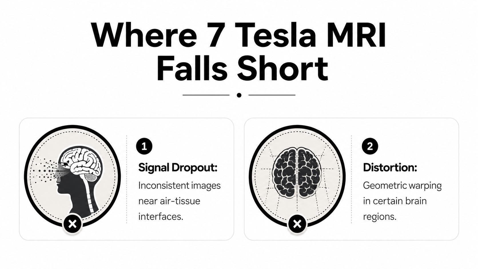
The biggest myth about 7 tesla MRI is that higher field strength always means a better answer. In practice, ultra-high-field imaging can lose reliability in certain anatomic regions because of the way physics and tissue properties interact. A technical review notes signal dropout and distortion at the skull base, including the orbitofrontal and inferior temporal regions, and describes similar signal loss in the cerebellum when B0 and B1+ inhomogeneity and higher SAR interfere with image quality.
For oncology, that limitation is not theoretical. Many important lesions sit near air-tissue interfaces or other difficult boundaries, where the scan can become harder to read instead of easier. If the area of concern is one of those regions, a 7T study may be less practical than a well-performed 3T exam.
The limits that matter most in cancer care
Outside the brain, the main problem is that body imaging at 7T remains challenging. That makes it a poor fit for many abdominal, pelvic, and thoracic oncology questions, where standard MRI protocols are already built to manage motion, depth, and larger anatomy.
Clinical coverage also makes clear that 7T is case-dependent, not universal. The same body of literature describes use in neurodegenerative disease, cerebrovascular disease, and ALS, but still mainly in specialized centers rather than routine community practice. For cancer care, that means the scanner alone does not determine value. Access, protocol design, and reader experience matter just as much.
Why this should change expectations
A better question is where 7T solves a problem that lower-field MRI cannot solve. That framing helps patients and oncologists separate genuine diagnostic gains from the assumption that stronger magnets automatically produce better care.
Clinical honesty helps here. If a 3T scan already answers the question well, 7T may add complexity without changing care.
That is especially true when treatment decisions depend on speed, comfort, and availability. A perfect image is not always the best clinical image if the patient cannot access it in time, and that applies just as much to the first oncology visit, which is why patients often benefit from a clear overview like what to expect at a first oncology appointment.
What to Expect During a 7 Tesla MRI Appointment
A patient arriving for a 7 tesla MRI should expect more screening than with a routine scan. The stronger magnetic field means the safety check is stricter, especially for implants, devices, prior surgeries, and anything metallic that could interfere with the scan or pose a risk. That first step is required, and it's one reason 7T appointments are usually handled at specialized centers.
The physical experience can feel different too. The scanner environment is still an MRI bore, but patients often notice that ultra-high-field systems are more intense in sound and feel. If you're prone to claustrophobia, that matters. The technologist's job is to keep the patient safe and still, and the radiologist's job is to make sure the protocol answers the clinical question without unnecessary sequences.
A typical flow
A typical visit starts with verification, then positioning, then the scan itself, followed by image review. Some patients will receive contrast if the ordering team thinks it's necessary, but the decision depends on the clinical target rather than the field strength alone. After the exam, a radiologist who understands ultra-high-field imaging interprets the data and sends a report back to the referring oncologist.
A few practical questions help patients prepare:
- Implants and devices: Tell the team about pacemakers, pumps, aneurysm clips, or any implanted hardware.
- Claustrophobia: Ask about the bore, communication options, and whether support measures are available.
- Noise: Expect a louder exam than many routine scans.
- Results timing: Ask who reads the images and how the report gets to the oncology team.
The most important thing is to ask whether the scan is meant to answer a very specific question. If the purpose is clear, the appointment feels less like a mystery and more like a targeted diagnostic step.
For patients who want a broader sense of what an oncology visit feels like, this first oncology appointment guide can help set expectations before the scan itself.
Accessing 7 Tesla MRI and Clinical Trials in New York City
In New York City, 7 tesla MRI is still a specialty resource, not a routine neighborhood test. Access is usually concentrated at major academic medical centers and research institutions, where the scanners, technologists, and radiologists are set up for ultra-high-field work. For most patients, that means the path starts with a treating oncologist or subspecialist who knows why the scan is being requested.
The next question is whether the result will help with a trial, a treatment decision, or both. Some clinical trials, especially in neuro-oncology and other specialized areas, use advanced imaging as part of eligibility screening or as an endpoint. When that's the case, the scan may help determine whether a patient fits a protocol or whether a lesion meets the trial's imaging criteria.
What patients should ask in NYC
A good referral conversation should cover a few basics.
- Why 7T instead of 3T: The ordering team should name the exact clinical question.
- Where the scan will be read: Ultra-high-field imaging should be interpreted by someone comfortable with its artifacts and strengths.
- How it fits the treatment plan: The result should connect to a real management decision.
- Whether a trial is involved: Imaging can sometimes support eligibility or trial monitoring.
The broader opportunity is coordination. In advanced cancer care, patients often need imaging, systemic therapy, and trial review to move in parallel. A specialty oncology practice can help make sure the scan does not sit in isolation, but instead feeds directly into the larger treatment plan.
For patients exploring research options, this clinical trials overview is a useful starting point for understanding how imaging and eligibility can intersect.
Why referral timing matters
If 7T is being considered, timing matters more than hype. The scan should be ordered when the clinical question is sharp enough to justify the trip, the protocol, and the interpretation. That is especially relevant for patients with treatment-resistant disease, where a subtle imaging detail may shape the next line of care.
Making Informed Decisions About Advanced Imaging
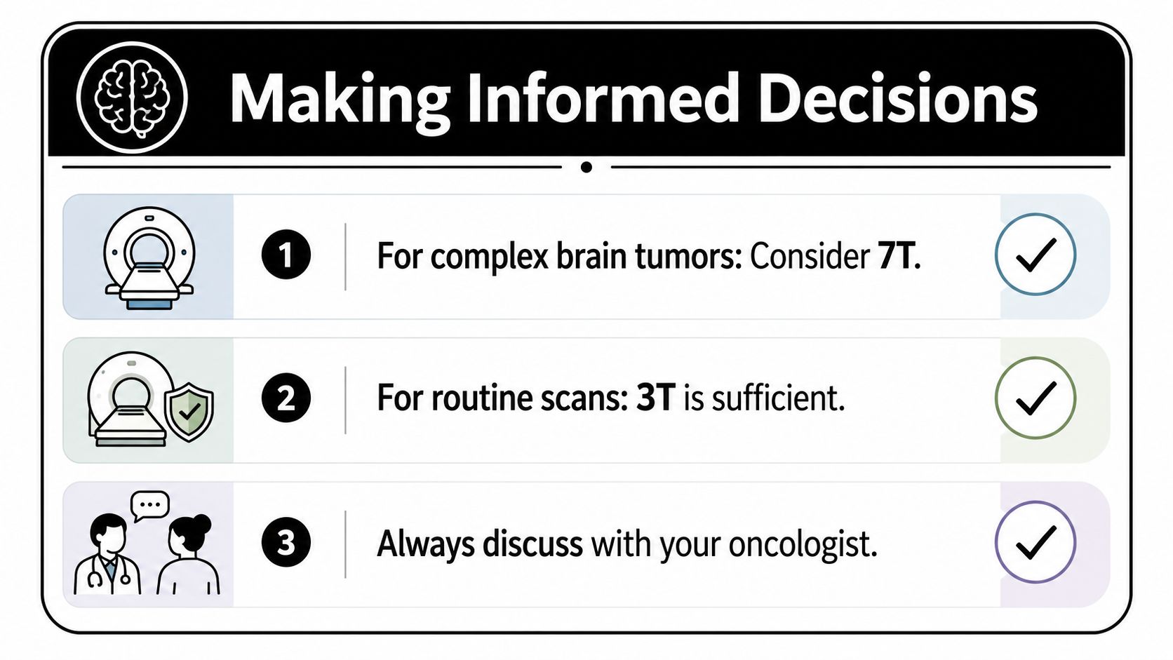
The best use of 7 tesla MRI is narrow but important. It shines when the question is about a tiny brain lesion, a subtle abnormality after treatment, or a trial-related imaging issue that needs maximum detail. It is much less compelling when the scan is routine, broad, or outside the brain, where 3T MRI often remains the more practical and reliable option.
A sensible decision framework looks like this.
- Complex brain lesions: Consider 7T when the clinical question depends on very fine anatomical detail.
- Routine oncology imaging: 3T is usually enough when the goal is standard staging or follow-up.
- Shared decision-making: The oncologist, radiologist, and patient should agree on what the scan needs to answer.
The bottom line for patients and clinicians
Advanced imaging works best when it is tied to a decision. If 7T will change the interpretation of a lesion, support a trial, or clarify a neurologic question, it deserves serious consideration. If it won't change management, the simpler scan is often the better scan.
The most informed patients are not the ones who chase the newest machine. They're the ones who ask, What will this scan change? That question keeps imaging grounded in care, not in hype.
If you're weighing advanced imaging as part of a complex cancer plan, the team at Hirschfeld Oncology can help you think through where specialized scans fit, how they connect to treatment options, and whether a referral pathway makes sense for your situation. Their Brooklyn practice works with patients facing advanced and treatment-resistant disease, and they can help you explore next-step questions with a care team that understands both the science and the stakes.
.png)
.png)