.png)


You're here because the symptoms didn't add up neatly. Maybe it started with tiredness that didn't improve, itching that seemed too minor for a specialist visit, or a yellow tint in the eyes that came and went. By the time someone says “we should look at the bile ducts,” the workup already feels slow, and the uncertainty can be exhausting.
Bile duct cancer diagnosis is hard for exactly that reason. The disease is rare, often subtle at first, and usually found only after several tests narrow the possibilities enough to justify tissue sampling and staging.
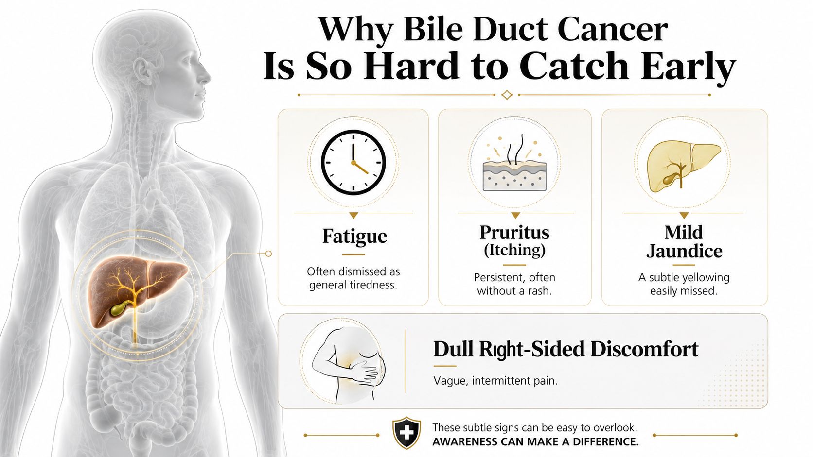
Why Bile Duct Cancer Is So Hard to Catch Early
A patient often comes in thinking the problem is something common. Fatigue feels like stress, itching feels like dry skin, mild jaundice can be blamed on gallstones or a liver flare, and dull right-sided discomfort is easy to dismiss as reflux or a muscular strain. That is how bile duct cancer stays hidden long enough to frustrate both patients and clinicians.
The rarity matters too. In the United States, about 8,000 people are diagnosed each year, and the American Cancer Society notes the true number may be higher because these cancers can be misclassified as something else when the first clues are unclear (American Cancer Society key statistics). Population registry data also show how uncommon the disease is, with intrahepatic bile duct cancer at 1.49 per 100,000 people and extrahepatic bile duct cancer at 0.96 per 100,000 during 2013 to 2017.
Why the first clue is often not dramatic
The diagnosis is hard partly because the earliest symptoms do not announce themselves as cancer. People usually feel unwell in a vague way first, then the labs or imaging create enough concern to keep going. In that sense, the process is less like flipping a switch and more like slowly turning down the uncertainty.
A useful mental model is a funnel. At the top are common explanations, such as skin irritation, gallstones, or a temporary liver problem. Each test narrows the list a little more, but early bile duct cancer can still sit in the narrow gap between a benign explanation and a clear answer. That is why doctors have to keep moving step by step, rather than relying on a single clue.
The low survival figures matter diagnostically as well as emotionally. The most recent data for people diagnosed between 2015 and 2021 show 5-year relative survival of 10% for intrahepatic bile duct cancer and 13% for extrahepatic bile duct cancer overall. Those numbers reflect how often the disease is found after it has already moved beyond the earliest, most treatable stage, which is exactly why the workup has to be careful and systematic.
Practical rule: if symptoms are persistent and the basic explanation does not fit, clinicians should keep widening the lens instead of settling too early on the first common diagnosis.
Symptoms and First Tests That Trigger a Workup
The first job is to decide whether the symptom pattern needs urgent attention. Unexplained jaundice, ongoing itching without a rash, dark urine, pale stools, or a persistent sense that something is “off” should prompt a medical visit, especially if the yellowing is new. A dull ache under the ribs or chronic fatigue alone may still turn out to be something benign, but when those symptoms cluster together, the odds shift enough to justify a fuller workup.
Blood tests usually come first. Doctors look at bilirubin, alkaline phosphatase, liver enzymes, and sometimes CA 19-9, not because any one value proves cancer, but because the pattern can suggest biliary obstruction or inflammation that needs imaging. A blood panel can tell the team that the liver and bile system need a closer look, yet it cannot tell them whether the cause is a stone, a stricture, cholangitis, or cancer.
What the first scan can and cannot do
An abdominal ultrasound is often the first image because it is fast and accessible. It works as a triage tool, helping clinicians see whether the bile ducts are dilated or whether there's a mass effect that needs more detailed imaging. It is useful, but it is not the finish line.
If the ultrasound is abnormal, or if jaundice remains unexplained, referral usually escalates to a specialist who can order cross-sectional imaging and decide whether tissue is needed. That escalation is the important step, because the diagnosis rarely rests on a single clean answer from one test. It is built by stacking clues until the probability becomes strong enough to justify the next move.
A negative first test doesn't always end the story. In biliary disease, the question is often whether the workup has lowered suspicion enough to stop, or whether it only moved the uncertainty into a more specialized phase.
The Imaging Ladder From Ultrasound to MRI
A patient may come in with jaundice, vague upper abdominal discomfort, or an abnormal lab pattern, and the first scan rarely settles the question by itself. Imaging works like a ladder because each step adds a little more certainty. Ultrasound is usually the first look, contrast-enhanced CT gives a wider anatomic view, and MRI with MRCP often becomes the clearest map of the biliary tree when clinicians need to understand ductal anatomy and the extent of the lesion, as outlined in a diagnostic workflow review. None of these tests alone gives the final answer, but each one lowers the number of possibilities.
MRI with MRCP is often favored because it shows the biliary system in high contrast and from multiple planes, which helps define parenchymal, biliary, and vascular extension. CT still matters when the team wants a broad look at tumor size, location, and nearby abdominal structures. Ultrasound remains useful as the starting point, but it is more of a sketch than a map. When clinicians are comparing cross-sectional imaging choices, a plain-language overview such as CT scan vs MRI on Hirschfeld Oncology can help frame the trade-offs.

How clinicians choose the next image
The next test depends on the question that remains open. If the main concern is whether the ducts are blocked, ultrasound may be enough to justify moving on. If the team needs a clearer look at the liver and surrounding structures, CT becomes more useful. If the key problem is mapping the ducts precisely enough to plan drainage, surgery, or sampling, MRI/MRCP often becomes the preferred study.
That preference does not make MRI definitive. Imaging narrows the field of possibilities, but it rarely gives the final answer on its own, and the review notes that imaging alone cannot reliably establish final pathology or fully settle local extent in every case (diagnostic workflow review). In practice, the scan with the most detail is the one that helps the team decide where uncertainty has dropped enough to move forward, and where it has not.
Ultrasound answers whether there is a problem worth pursuing. CT shows where the abnormality sits and what else may be involved. MRI/MRCP shows how the biliary anatomy is arranged. None of those questions is the same as asking whether this is cancer, which is why the workup continues when suspicion remains.
For patients, the sequence is less like a single verdict and more like assembling a medical map piece by piece. Each study makes the next choice clearer. It also exposes the limits of early detection, because small or subtle bile duct tumors can still hide in plain sight until the anatomy has been examined from several angles.
Why the sequence matters
The ladder matters because different scans answer different questions, and the order changes what the team can see. A quick ultrasound can show dilation or a blocked duct, but it cannot always trace the cause. CT adds a broader view of the liver, pancreas, vessels, and other abdominal organs. MRI with MRCP adds detail about the ducts themselves, which is often the part clinicians need most when they are trying to understand a suspected cholangiocarcinoma.
That is why the diagnostic journey usually feels gradual rather than abrupt. Each step trims away some uncertainty, yet early disease can remain difficult to catch because imaging is still limited by small lesion size, overlapping inflammation, and the complexity of the bile ducts. The process is built around probability, not certainty, and the image that looks most convincing still has to fit with the rest of the clinical picture.
Choosing Between ERCP and EUS for Tissue Diagnosis
A patient may come away from imaging with a clear concern but no tissue proof yet. That is usually the point where the team decides how to get a sample without wasting time or missing the best target. In biliary disease, ERCP and EUS answer different questions. One looks into the duct and can help relieve blockage. The other samples from outside the duct and often sees nearby nodes or a hidden mass more clearly.
ERCP is often chosen when a stricture needs both diagnosis and drainage. During the same procedure, the physician can collect brush cytology, take a biopsy, use FISH analysis, and sample under cholangioscopy, so the procedure can serve more than one purpose at once. That matters when jaundice is present and the team also needs to plan for biliary stent placement. EUS is often a better fit when the concern is a mass beside the duct, local invasion, or nearby lymph nodes, because it can sample from the stomach or duodenum without entering the bile duct itself.
ERCP vs EUS for Bile Duct Cancer Tissue Diagnosis
| Feature | ERCP | EUS |
|---|---|---|
| Main use | Samples a duct stricture and can relieve blockage | Samples a mass, nearby nodes, or local extension |
| Tissue methods | Brush cytology, biopsy, FISH, cholangioscopy-guided sampling | Fine-needle sampling of masses or nodes |
| Strength | Can combine diagnosis and drainage in one session | Strong for lesions outside the duct and staging nearby spread |
| Trade-off | Involves instrumentation of the bile duct | Does not directly address duct drainage in most cases |
| When it helps most | Obstructive jaundice with a duct lesion that may need a stent | Suspected mass or nodes where duct access is not the main issue |
The choice is less about which test is better in general and more about which test answers the next question with the least detour. If the duct is blocked and the patient is jaundiced, ERCP may solve the sampling problem and the drainage problem together. If the concern is a mass or nodal spread, EUS may give the cleaner picture of what tissue is involved and how far the process extends.
Recovery and risk are part of the trade-off too. ERCP can cause pancreatitis, bleeding, and infection, while EUS has its own procedural considerations and is not a drainage procedure in most cases. That is why the plan is often built around the anatomy in front of the team, not around a fixed rule. A sample is only useful if it is taken from the place most likely to change the diagnosis or the treatment path.
If biopsy tools are being prepared, many endoscopy units also rely on precise sampling supplies, including sterile punch medical supplies, because tissue quality can matter as much as the procedure itself.
Staging and Molecular Profiling That Shape Treatment
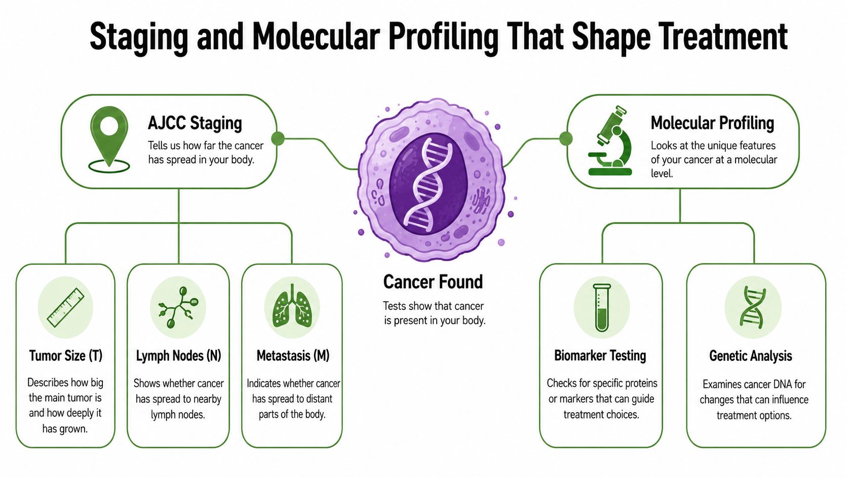
Once cancer is confirmed, the next question is not just “what is it?” but “how far has it gone, and what makes this tumor different from the others?”. Staging describes the size and spread of the cancer, while molecular profiling looks for features inside the tumor that can change treatment options. Together, they turn a diagnosis into a map for decision-making.
The AJCC system organizes staging around the familiar T, N, and M framework. T refers to the primary tumor, N to lymph nodes, and M to distant spread. Imaging and pathology are combined to assign that stage, because neither one tells the whole story by itself.
Why biomarker testing changes the conversation
Molecular profiling may include NGS, FISH, and immunohistochemistry to look for alterations such as FGFR2 fusions, IDH1/2 mutations, HER2 amplification, BRAF V600E, and MSI-H/dMMR. Those markers matter because they can open the door to targeted therapy or a clinical trial when standard chemotherapy offers limited effectiveness. For some patients, that result changes the plan from a general regimen to a far more specific one.
A single biomarker finding can redirect the entire path. For example, if profiling identifies an actionable target, the oncology team may prioritize a targeted agent or a trial instead of relying only on conventional treatment. If the testing is unrevealing, that doesn't mean the work was wasted, it means the team has ruled out one more avenue and can focus on the best remaining options.
For a deeper look at how tumor testing works in practice, Hirschfeld Oncology also has a clear overview of molecular testing for cancer.
Clinical insight: a precise diagnosis doesn't just name the cancer. It can determine whether the next step is surgery, drainage, targeted therapy, immunotherapy, or a trial the patient wouldn't otherwise know to ask about.
Why Diagnosis Is Often a Probability, Not a Verdict
Patients often expect a scan or biopsy to end the uncertainty. In bile duct cancer, that's not always how it works. A brush cytology can come back negative even when suspicion remains high, and an inflammatory stricture can look malignant on imaging because inflammation and cancer can overlap so closely.
That's why multidisciplinary review matters. Surgeons, gastroenterologists, radiologists, pathologists, and oncologists may all look at the same case and still disagree on the level of certainty. When the lesion is small or risky to biopsy, the team may choose repeat testing, short-interval follow-up, or additional sampling rather than pretending the first answer is final.
Living with an incomplete answer
The hard part for patients is that uncertainty itself becomes part of the diagnosis. A report may say “suspicious,” “indeterminate,” or “concerning for malignancy,” and those words are not failure, they're often the honest reflection of what the evidence can support at that moment. That is especially true in biliary disease, where the first test may only change the odds, not settle the question.
Second opinions can help when the path isn't clear. They're not about distrust, they're about making sure a rare and technically difficult diagnosis gets seen by a team that deals with it often. Watchful waiting can also be appropriate in select situations, but only when the specialist can explain exactly what would trigger the next step and why.
Questions to Ask at Your Specialist Consultation
Bring a written list. In a complex bile duct cancer diagnosis, patients do better when the next decision is framed in plain language instead of scattered across several appointments. If you're heading to a hepatobiliary surgeon, gastroenterologist, or medical oncologist, ask the questions that force the team to define the diagnosis and the plan clearly.
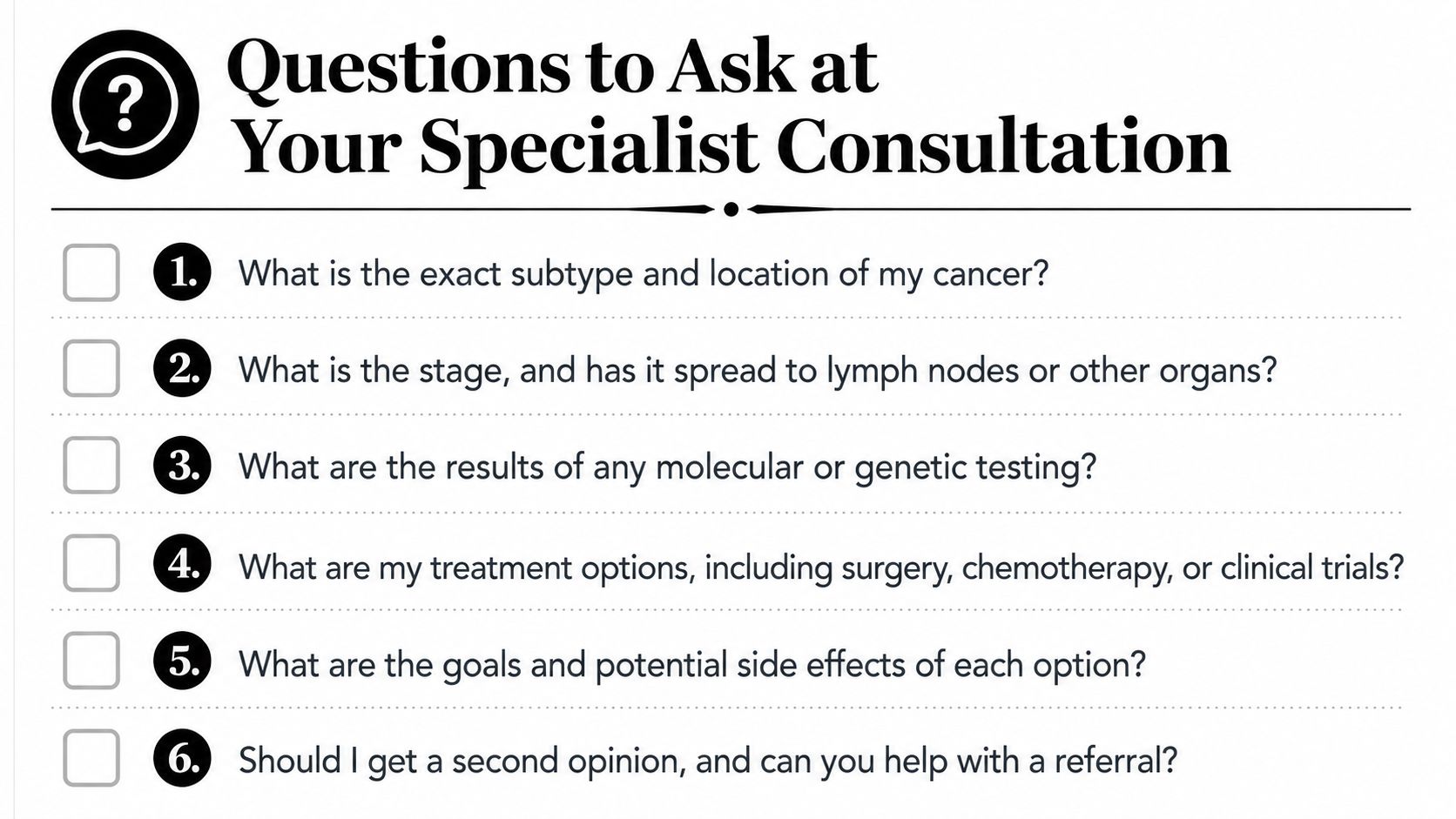
The questions that move care forward
- What is the exact subtype and location of my cancer? That answer matters because intrahepatic and extrahepatic disease behave differently and may need different specialists.
- What is the stage, and has it spread to lymph nodes or other organs? This sets expectations for surgery, drainage, and systemic treatment.
- What are the results of molecular or genetic testing? If the report hasn't been done yet, ask when it will be sent.
- What are my treatment options, including surgery, chemotherapy, or clinical trials? Not every option will fit every stage.
- What are the goals and potential side effects of each option? Knowing the aim of treatment helps patients decide what trade-offs are acceptable.
- Should I get a second opinion, and can you help with a referral? For a rare cancer, that question is completely reasonable.
What to bring and what to ask about access
Bring imaging disks, pathology reports, procedure notes, and a list of all prior procedures. If the center is far from home, ask whether some infusion-based care can be coordinated closer to you through a community oncology partner. Hirschfeld Oncology, for example, is a Brooklyn practice and infusion center that sees bile duct cancer among its focused disease areas, which can matter for patients in neighborhoods like Williamsburg or Bushwick who need ongoing treatment without losing specialist oversight.
Practical rule: if the consultation ends without a clear subtype, stage, and next step, ask for the missing piece before you leave.
Turning a Diagnosis Into a Treatment Path
A difficult diagnosis still has value when it is accurate and complete. In bile duct cancer, the workup is usually multimodal, sometimes probabilistic, and often emotionally draining, but it gives the team enough information to make real treatment decisions instead of guessing. That's the point where care becomes specific, and specificity is what opens options.
The best teams are comfortable with complex disease, targeted therapy, immunotherapy, and carefully selected chemotherapy when standard approaches aren't enough. They also know when to pause, get another look, or coordinate a second opinion rather than rushing past the uncertainty. A clear diagnosis doesn't erase the burden, but it does give patients and clinicians something concrete to work with.
If you're facing a bile duct cancer workup, Hirschfeld Oncology can help you sort through staging, tissue diagnosis, and treatment planning in one coordinated oncology setting. Visit Hirschfeld Oncology to learn how the practice approaches complex cancers and to request a consultation when you're ready to discuss the next step.
.png)
.png)